| Brief communication | Peer reviewed |
Cite as: Di Provvido A, Trachtman AR, Farina E, et al. Pleurisy evaluation on the parietal pleura:An alternative scoring method in slaughtered pigs. J Swine Health Prod. 2019;27(6):312-316.
Also available as a PDF.
SummaryThe present study aims to develop and assess an alternative method for scoring pleurisy in slaughtered pigs. Overall, data indicates that pleurisy can be scored effectively and efficiently by inspecting the parietal pleura. Moreover, this evaluation can be suitably carried out on digital images, thus optimizing the workload of veterinarians. | ResumenEl objetivo del presente estudio es desarrollar y evaluar un método alternativo de puntuación de pleuresía en cerdos sacrificados. En general, la información indica que la pleuresía puede evaluarse efectiva y eficientemente mediante la inspección de la pleura parietal. Además, esta evaluación puede hacerse apropiadamente en imágenes digitales, optimizando así la carga de trabajo de los veterinarios. | ResuméLa présente étude visait à développer et évaluer une méthode alternative de noter la pleurésie chez des porcs abattus. Globalement, les données indiquèrent que la pleurésie peut être notée de manière efficace et compétente en inspectant la plèvre pariétale. De plus, cette évaluation peut être effectuée adéquatement sur des images digitales, optimisant ainsi la charge de travail des vétérinaires. |
Keywords: swine, slaughterhouse, pleurisy, scoring methods
Search the AASV web site
for pages with similar keywords.
Received: March 18, 2019
Accepted: May 15, 2019
The slaughterhouse is recognized worldwide as a useful check point for assessing the health status of livestock, as well as the effectiveness of strategies implemented to prevent or treat disease conditions. This is especially true for pigs, since their lifespan does not permit the full healing of lesions, which are often still evident at slaughter.1-4
Several methods have been developed to quantify the impact of diseases. However, the greatest attention has always been paid to the so-called porcine respiratory disease complex, which deeply reduces the profitability of pig farming.3-5
Regardless of the animal species and the disease taken into consideration, each scoring method should fit some general requirements: a) it should be simple, fast, and compatible with the slaughter line speed; b) it should be easily standardizable and reproducible; and c) it should provide data that can be easily interpreted and analyzed.3
Pleurisy is commonly observed at necropsy or during the postmortem inspection at the abattoir, its prevalence often being close to or above 50% in slaughtered pigs.2 Multiple pleurisy scoring systems have been developed over the years; among these is the slaughterhouse pleurisy evaluation system grid (SPES),6 which suitably meets the above criteria and is widely used to quantify pleurisy caused by Actinobacillus pleuropneumoniae (App) infection.2,3,7
The present work aims to assess an alternative method to score pleurisy in slaughtered pigs, based on the inspection of the parietal pleura. This method has been compared with the SPES grid, which is considered the gold standard in this field of study. The feasibility of scoring pleurisy on digital images has also been thoroughly examined.
Materials and methods
Animals
A total of 476 heavy pigs (9-11 months of age; 150-180 kg) were included in the present study. These pigs were slaughtered in Central and Northern Italy, between November 2017 and June 2018. The study was performed in 2 distinct steps, scoring pleurisy at the slaughterhouse and using digital images.
Scoring pleurisy at the slaughterhouse
Two hundred sixteen slaughtered pigs were investigated. The scoring was carried out by 3 skilled veterinary surgeons after a training period and reaching consensus about how to score and record lesions. Specifically, the presence or absence and the features of pleurisy were evaluated.
A veterinarian was stationed on the slaughter line where the postmortem inspection of viscera is usually performed. The inflammatory reaction of the visceral pleura (ie, the serous membrane lining the lungs) was scored according to the SPES grid (Table 1) and reported in an ad hoc format.6 Another veterinarian was at a different point of the slaughter line and inspected the parietal pleura (ie, the serous membrane lining the chest wall). The presence or absence of pleurisy was reported in an ad hoc format and scored using the pleurisy evaluation on parietal pleura (PEPP) scale as detailed in Figure 1. Pleurisy was scored in each area regardless of the extent of lesions in order to limit the subjectivity of the judgment. According to the SPES grid,6 considering the topography of the thoracic organs and that App-induced lesions usually affect the diaphragmatic lung lobes,8 the following scores were established: 1 point for pleurisy affecting the cranial area of the parietal pleura; 2 points for pleurisy affecting the middle area of the parietal pleura; 3 points for pleurisy affecting the remaining caudal area of the parietal pleura. The points of both carcass halves are summed for a total score for each pig ranging from 0 to 12 (explanatory examples are shown in Figure 2).
Table 1: Scoring pleurisy by the SPES method6
| Score | Features of pleurisy, considering the extension and localization of lesions |
|---|---|
| 0 | Absence of lesion. |
| 1 | Pleurisy affecting the cranial-ventral portion of the lung; interlobar adhesion. |
| 2 | Discrete, unilateral pleurisy of the diaphragmatic lobe. |
| 3 | Discrete, bilateral pleurisy of both the diaphragmatic lobes; large, unilateral adhesion affecting the diaphragmatic lobe. |
| 4 | Large, bilateral adhesions between both the diaphragmatic lobes on one side and the chest wall on the other. |
Figure 1: The parietal pleura was divided into three, easily identifiable areas: 1) from the 1st to the 3rd intercostal space; 2) from the 4th to the 6th intercostal space; and 3) all the remaining caudal intercostal spaces.
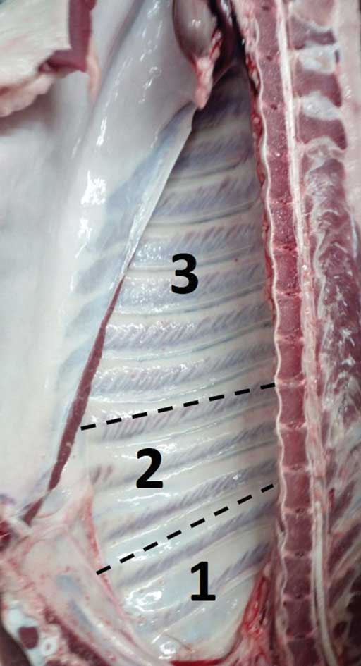
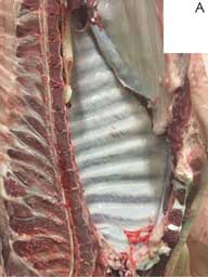
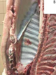
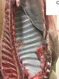
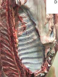
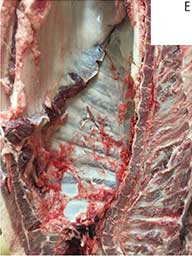
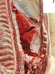
Figure 2: Scoring pleurisy using the PEPP method. A) Pleurisy affecting the cranial portion of the chest wall, corresponding to a score of 1. B) Pleurisy of the 4th and 5th intercostal spaces, corresponding to a score of 2. C) Pleurisy affecting both the cranial and the middle portion of the chest wall, corresponding to a total score of 3. D) Pleurisy affecting the caudal intercostal spaces, corresponding to a score of 3. E and F) The entire parietal pleura was affected by pleurisy, corresponding to a total score of 6. The points of both carcass halves are summed for a total score for each pig ranging from 0 to 12. PEPP = pleurisy evaluation on parietal pleura.
Scoring pleurisy using digital images
The reliability of scoring pleurisy on digital images was also evaluated. A veterinarian scored lesions at the slaughterhouse using the PEPP method and took pictures of all the animals under study (n = 260). These pictures were shared with 2 veterinarians, who independently applied the PEPP method, being unaware of the score given at the slaughterhouse.
Statistical analysis
The suitability of the sample size was assessed for a generalized linear model using G* Power.9 The mean scores obtained by applying SPES and PEPP were compared according to the diagnostic outcome (negative vs positive) by one-way analysis of variance. The relationship between the scores obtained with the 2 methods was evaluated using the Pearson’s linear correlation coefficient (r). The functional relationship between the variables measured with the 2 scoring methods was solved by linear regression analysis, whose statistical significance was evaluated by the analysis of variance; the appropriateness of the fitting was estimated using the coefficient of determination (R2).
The correlation among the scores obtained by applying the PEPP method at the slaughterhouse and on digital pictures was investigated and expressed by the Pearson’s linear correlation coefficient (r). Finally, the agreement between the 2 veterinarians scoring pleurisy on digital images was measured by the Cohen’s kappa coefficient (κ value).
Results
Scoring pleurisy at the slaughterhouse by SPES and PEPP methods
The presence of visceral pleurisy was demonstrated in 109 of 216 pigs (50.46%), while no pleural inflammation was detected in the remaining 107 of 216 pigs (49.54%) by the application of the SPES grid. On the same pigs, the application of the PEPP method demonstrated the presence of inflammatory reactions of the parietal pleura in 108 of 216 pigs (50%), while the remaining 108 of 216 pigs (50%) were considered healthy.
The scores obtained using both SPES and PEPP are shown in Figure 3. The similarity between the 2 scoring systems appears quite evident, although based on a different reference scale. In particular, the total number of healthy pigs (score 0) was almost identical. Actually, 8 pigs showing interlobar adhesions (score 1 with the SPES grid) were erroneously regarded as healthy by applying the PEPP method; on the other hand, 7 pigs with small lesions affecting the cranial intercostal spaces (score 1 with the PEPP method) were missed by applying the SPES grid.
Figure 3: Pleurisy scores obtained by applying the A) SPES and B) PEPP pleurisy evaluation systems. Approximately, 50% of the pigs evaluated showed no pleural lesion and obtained a score of 0 using both scoring systems. The distribution of scores using the SPES system was rather uniform, with a score of 2 most frequently recorded. Using the PEPP method, most of the pigs with pleural lesions received scores of 1 to 6, with a small number of pigs scoring > 7. SPES = slaughterhouse pleurisy evaluation system; PEPP = pleurisy evaluation on parietal pleura.
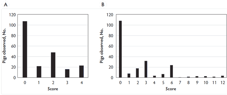
Overall, the PEPP method was able to effectively discriminate diseased from healthy pigs (P < .001), when compared with the SPES grid. The scores obtained with the 2 methods showed a very high Pearson’s correlation coefficient (r = 0.913), which was statistically significant (P < .001). The linear regression analysis indicated that the coefficient of determination was very high (R2 = 0.833) and statistically significant (P < .001).
Scoring pleurisy using digital images
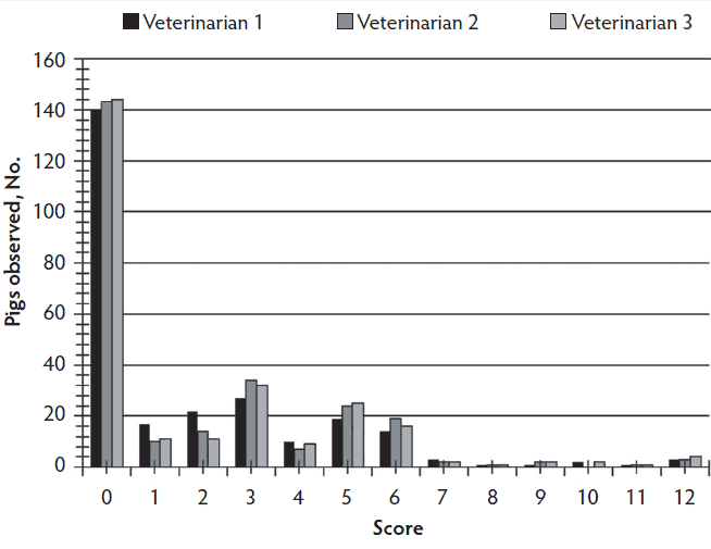
Scoring lesions on digital images proved to be quite easy and fast (around 8 pigs/minute, including recording scores in a spreadsheet). The scores obtained using the PEPP method at the abattoir and on digital images are shown in Figure 4, which underlines the high level of similarity among independent investigators. In particular, the number of healthy pigs (score 0) was almost identical, ranging between 140 to 144 of 260 pigs. The correlation among the investigators proved to be very high and statistically significant (r = 0.89 and 0.94; P < .001). Finally, the agreement between the 2 veterinarians scoring pleurisy on digital images was also very high (κ value = 0.852).
Figure 4: Pleurisy scores obtained by applying the PEPP method to carcasses at slaughter (veterinarian 1) and to digital pictures of the carcasses (veterinarians 2 and 3). PEPP = pleurisy evaluation on parietal pleura.
Discussion
The examination of slaughtered pigs is extremely useful and cost-effective to assess the health status of livestock, along with data collected in the herds (eg, clinical signs, necropsy findings, consumption of drugs, daily weight gain, and feed efficiency) or resulting from laboratory tests (eg, serological surveys).3,5 For this reason, the assessment of innovative and suitable scoring methods is always highly desirable.
Actinobacillus pleuropneumoniae is the etiologic agent of porcine pleuropneumonia, a respiratory disorder of pigs distributed worldwide, causing significant economic losses to the swine industry.8 A large body of evidence indicates that a high prevalence of chronic adhesive pleuritis at slaughter is very suggestive of previous App infection, thus further emphasizing the importance of the abattoir as a valuable source of data.2,5,10,11 Different scoring systems have proven suitable to quantify App lesions. However, the SPES grid is the only one that can be reliably assessed under field conditions, hence why it is considered the most informative system worldwide.7
Overall, our data indicate that the SPES and PEPP methods provide well matching results. We consider this to be widely expected, as pleurisy usually involves both pleural sheets (visceral and parietal), with very rare exceptions being possible (eg, interlobar pleuritis). Therefore, the PEPP scoring method could represent a reliable alternative to the SPES grid.
Obviously the PEPP method, like all the others, shows both strengths and weaknesses. For example, the inspection of the parietal pleura may not be compatible with the simultaneous evaluation of other lesions (eg, pneumonia, pericarditis, parasitic hepatitis). On the other hand, the PEPP method seems to be simple, not very influenced by possible confounding factors (eg, blood staining, lung scarring), and it can be applied in alternate locations on or off the slaughter line. In addition, it could be much faster than other methods if all carcasses are available at the end of the slaughter chain.
In our opinion, the effective application of the PEPP method on digital images could be particularly useful. The same approach appears difficult if not impossible for the SPES method because of a number of practical issues: (a) the difficulty in obtaining good quality images of the lungs along the slaughter chain; (b) the presence of large amounts of blood on the surface of viscera, including lungs; and (c) the inspiration of blood and water into the lungs from the scalding tank. Our data indicate that scoring pleurisy on digital pictures of the chest wall is fast, relatively simple, and easily standardizable, providing results which are largely comparable with those obtained by a veterinarian at the slaughterhouse. Therefore, this could be timesaving, efficient, and effective, notably streamlining the workload of the investigators.
Implications
- Pleurisy evaluation of parietal pleura was effective and efficient.
- Using PEPP on digital images was effective and optimized inspector time.
Acknowledgments
The present study has been carried out in the framework of the Project “Demetra” (Dipartimenti di Eccellenza 2018-2022, CUP_C46C18000530001), funded by the Italian Ministry for Education, University and Research. The authors gratefully thank Mr Andrea Paolini for his kind collaboration in collecting digital pictures at the slaughterhouse.
Conflict of interest
None reported.
Disclaimer
Scientific manuscripts published in the Journal of Swine Health and Production are peer reviewed. However, information on medications, feed, and management techniques may be specific to the research or commercial situation presented in the manuscript. It is the responsibility of the reader to use information responsibly and in accordance with the rules and regulations governing research or the practice of veterinary medicine in their country or region.
References
1. Sanchez-Vazquez MJ, Strachan WD, Armstrong D, Nielen M, Gunn GJ. The British pig health schemes: integrated systems for large-scale pig abattoir lesion monitoring. Vet Rec. 2011;169:413.
2. Merialdi G, Dottori M, Bonilauri P, Luppi A, Gozio S, Pozzi P, Spaggiari B, Martelli P. Survey of pleuritis and pulmonary lesions in pigs at abattoir with a focus on the extent of the condition and herd risk factors. Vet J. 2012;193:234-239.
3. Luppi A, Merialdi G. Lesioni al macello [Slaughter injuries]. In: Martelli, P, ed. Le Patologie del Maiale [The Pathologies of the Pig]. Milan, Italy: Point Veterinaire Italie; 2013:199-218.
4. Scollo A, Gottardo F, Contiero B, Mazzoni C, Leneveu P, Edwards SA. Benchmarking of pluck lesions at slaughter as a health monitoring tool for pigs slaughtered at 170kg (heavy pigs). Prev Vet Med. 2017;144:20-28.
5. VanAlstine WG. Respiratory System. In: Zimmerman JJ, Karriker LA, Ramirez A, Schwartz KJ, Stevenson GW, eds. Diseases of Swine. 10th ed. Ames, IA: Wiley-Blackwell Publishing; 2012:348-362.
6. Dottori M, Nigrelli AD, Bonilauri P, Merialdi G, Gozio S, Cominotti F. Proposta per un nuovo sistema di punteggiatura delle pleuriti suine in sede di macellazione: la griglia SPES (Slaughterhouse Pleurisy Evaluation System). Large Anim Rev. 2007;13:161-165.
7. Sibila M, Aragón V, Fraile L, Segalés J. Comparison of four lung scoring systems for the assessment of the pathological outcomes derived from Actinobacillus pleuropneumoniae experimental infections. BMC Vet Res. 2014;10:165. doi:10.1186/1746-6148-10-165
8. Gottschalk M. Actinobacillosis. In: Zimmerman JJ, Karriker LA, Ramirez A, Schwartz KJ, Stevenson GW, eds. Diseases of Swine. 10th ed. Ames, IA: Wiley-Blackwell Publishing; 2012:653-669.
9. Faul F, Erdfelder E, Lang AG, Buchner A. G*Power 3: a flexible statistical power analysis program for the social, behavioral, and biomedical sciences. Behav Res Methods. 2007;39:175-191.
10. Fraile L, Alegre A, López-Jiménez R, Nofrarías M, Segalés J. Risk factors associated with pleuritis and cranio-ventral pulmonary consolidation in slaughter-aged pigs. Vet J. 2010;184:326-333.
11. Meyns T, Van Steelant J, Rolly E, Dewulf J, Haesebrouck F, Maes D. A cross-sectional study of risk factors associated with pulmonary lesions in pigs at slaughter. Vet J. 2011;187:388-392.