| Brief communication | Peer reviewed |
Cite as: Dewey C, Sunstrum J, Richardson K. Management of ear hematomas in pigs. J Swine Health Prod. 2018;26(3):137-141. https://doi.org/10.54846/jshap/1029
Also available as a PDF.
SummaryThis study compared two treatment options to manage ear hematomas and measured impact of hematomas on growth rate. Incised ears were more likely to become infected than those not incised but did not differ in time to resolution of the hematoma. Pigs with hematomas grew slower than those without hematomas. | ResumenEste estudio comparó dos opciones de tratamiento para manejar hematomas de oreja y medir el impacto de los hematomas en el índice de crecimiento. Las orejas cortadas fueron más propensas a infectarse que las nos cortadas pero no difirieron en el tiempo de resolución del hematoma. Los cerdos con hematomas crecieron más lento que los que no tuvieron hematomas. | ResuméLa présente étude visait à comparer deux options de traitement pour la gestion des hématomes d’oreille et à mesurer l’impact des hématomes sur le taux de croissance. Des oreilles incisées étaient plus susceptibles de s’infecter que celles non-incisées mais il n’y avait pas de différence dans le temps pour la résolution des hématomes. Les porcs avec des hématomes avaient une croissance ralentie comparativement à ceux sans hématome. |
Keywords: swine, ear, aural, hematoma, treatment
Search the AASV web site
for pages with similar keywords.
Received: February 15, 2017
Accepted: December 5, 2017
Aural (ear) hematomas occur due to the rupture of blood vessels resulting in a collection of blood predominantly in the subperichondral region of the cartilage within the pinna.1 Rupture of the blood vessels within the cartilage is typically due to physical trauma from violent shaking of the ear in response to sarcoptic mange or pediculosis, bites on the ears from other pigs, necrotic ear syndrome, from handling the pig by the ear, or injuries on barn equipment.2 If left untreated, a hematoma will resolve without any intervention; however, this can take several weeks and the hematoma may be of sufficient size as to reduce feed intake.1 To the authors’ knowledge, no research has been published that describes the best methods to treat hematomas as a means to optimize resolution of the hematoma and the growth rate of the pig.
In pot-bellied pigs, treatment options include leaving the hematoma alone to spontaneously open and drain, inserting an indwelling plastic teat tube, or surgical incision into the most fluctuant area of the hematoma.3 Similar to pot-bellied pigs, dogs can also develop ear hematomas and several treatment options are available. In dogs, treatment involves either medical management with corticosteroids, with or without concurrent draining of the hematoma, or surgical intervention.4 Surgical intervention most commonly involves either placement of a teat cannula, a closed suction drain, or an incision into the pinna to drain the hematoma and full thickness mattress sutures to compress the cavity.5 At least one source recommends that hematomas in commercial pigs be surgically repaired in a similar manner.2 The objectives of this study were to evaluate the impact of surgical intervention of the ear hematoma on resolution time; infection rate and severity; and growth rate 3 weeks post intervention.
Materials and methods
The protocol for this study was approved by the University of Guelph Animal Care Committee.
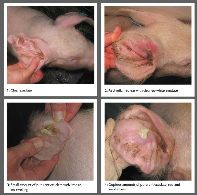
A convenience sample of four swine farms was selected to participate in this study. Farms were visited on a weekly basis for 7 to 18 weeks. During this time, all nursery pigs with ear hematomas were enrolled in the study and matched to negative-control pigs with similar weights. Pigs with hematomas were randomly assigned to one of two treatments: surgical incision or no intervention (positive control). On each farm, systematic random sampling was used such that the first three pigs identified with hematomas were randomly assigned to the surgical-incision group or positive-control group so that within each block of three pigs there were two in the surgically-incised group and one in the positive-control group. More pigs (66%) were assigned to the surgical-incision group because that was identified as the optimal treatment according to reference textbooks.2,6 Subsequent blocks of three pigs were assigned to groups in the same pattern as that of the first block. Pigs in the surgically-incised group had the skin of the ear incised to release the blood, but in the positive-control group the pig’s ear was left untreated. A uniquely numbered ear tag was put in the healthy ear. Upon enrollment, pigs were weighed and assigned swelling and infection scores. They were re-weighed and re-examined weekly for 3 weeks post identification. Swelling was scored on a scale of 0 to 4 where 0 indicated little to no swelling, 1 indicated slight swelling, 2 indicated moderate swelling, 3 indicated severe swelling, and 4 indicated swelling extended as much as the skin would accommodate (Figure 1). The hematoma was considered resolved if the swelling score was < 1 following the first, second, or third week after the pig entered the study. Infection was scored on a scale of 0 to 4 where 0 indicated no exudate, 1 indicated clear exudate, 2 indicated redness with clear or white exudate, 3 indicated a small amount of purulent exudate, and 4 indicated copious amounts of purulent exudate, redness, and swelling (Figure 2). On each farm, two pigs with approximately the same weight and in the same pen as the pig with the hematoma were selected for the negative-control group. These pigs were ear tagged and weighed upon enrollment and then weekly for 3 subsequent weeks. All pigs remained in their home pen as was the management practice of the farms included in the study.
Figure 1: Examples of swelling scores from 1-4 and a pig with a resolved hematoma.
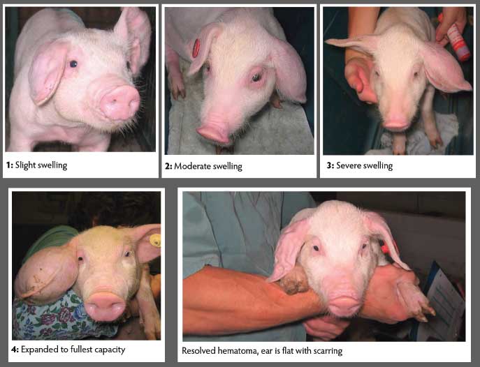
Figure 2: Examples of infection scores from 1-4 with 4 being the most infected.
Pigs in the negative-control group were removed prior to the analyses of the study if the weight upon enrollment differed from that of the matched pig (with the hematoma) by more than 1 kg. Pigs in the surgically-incised and positive-control groups were removed prior to the analyses if they did not have at least one weight-matched negative-control pig or if they died prior to being weighed 3 weeks after enrollment. There were 77, 47, and 213 pigs in the surgically-incised, positive-control, and negative-control groups respectively. A Wilcoxin rank sum test was used to compare the proportion of pigs whose hematomas were resolved by week between surgically-incised and positive-control groups. A Wilcoxin rank sum test was also used to compare the proportion of pigs with ear infections and the extent of the ear infection by week between surgically-incised and positive-control groups. The weight of the pig at each week was compared among treatment groups using an ANOVA after controlling for the farm of origin. The average daily gain (ADG) was calculated for the first-, second-, and third-week weights after the pigs were enrolled in the study. An ANOVA was used to determine the association between weekly ADG, the presence of a hematoma, and the treatment group after controlling for the weight of the pig at the start of the time period and the farm of origin. All weights and ADG were presented as least squares means after controlling for farm of origin. These statistics were calculated using Statistix version 9 software (Analytical Software, Tallahassee, Florida).
Results
The average weight of the pigs enrolled in the study did not differ by group (P > .05). At enrollment, pigs in the surgically-incised, positive- and negative-control groups weighed 11.58, 11.04, and 11.21 kg respectively (Table 1). Pigs in the surgically-incised group had a lower ADG in the first and second week after incising the hematoma than did pigs in the negative-control group (P < .01). Pigs in the surgically-incised group had a lower ADG in the first and second week after treatment than did the pigs in the positive-control group (P = .01). The ADG of pigs with hematomas did not differ from that of pigs without hematomas in the third week after enrollment. The ADG of pigs from enrollment until week 3 in the surgically-incised group was lower than that of the positive- and negative-control groups (P < .01). However, the increase in the ADG from the first week of the study to the last week of the study was larger in the surgically-incised group than in the other two groups, indicating that there was compensatory gain in the pigs in the treatment group (P = .001) (Table 1).
Table 1: Weight and average daily gain of pigs enrolled in a clinical trial to compare treatment options for hematomas
| No hematoma (Negative control) | Hematoma not incised (Positive control) | Surgically incised | P | ||||
|---|---|---|---|---|---|---|---|
| Mean | SE | Mean | SE | Mean | SE | ||
| Weight (kg)* | |||||||
| Week 0 | 11.58a | .261 | 11.04a | .469 | 11.21a | .386 | .47 |
| Week 1 | 14.20a | .291 | 13.53ab | .532 | 13.17b | .438 | .10 |
| Week 2 | 17.45a | .336 | 16.25ab | .604 | 16.02b | .497 | .02 |
| Week 3 | 21.98a | .385 | 19.46b | .691 | 19.45b | .569 | .02 |
| ADG (kg)† | |||||||
| Week 1 | .42a | .014 | .40a | .012 | .32b | .019 | < .001 |
| Week 2 | .51a | .017 | .49ab | .026 | .46b | .023 | .018 |
| Week 3 | .51a | .021 | .51a | .031 | .52a | .027 | .91 |
| Weeks 1 to 3 | .48a | .009 | .45ab | .018 | .43b | .017 | .01 |
| Increase week 1 to week 3 | .13a | .018 | .124a | .033 | .226b | .027 | .001 |
* ANOVA presenting least squares means after controlling for farm of origin.
† ANOVA presenting least squares means after controlling for farm of origin and weight of the pig at the start of the week.
ab Different superscripts within rows indicates significantly different mean values.
SE = standard error; ADG = average daily gain.
Pigs in the surgically-incised group were more likely to have infections and were more likely to have more severe infections in weeks 1, 2, and 3 after incising the ear than pigs in the positive-control group (P < .01) (Table 2). There were 47 pigs (61%) in the surgically-incised group whose ears became infected following treatment, whereas only three pigs (1.5%) in the positive-control group had infected ears. In the surgically-incised group, 14, seven, and three pigs had maximum infection scores with a severity of 2, 3, and 4, respectively, whereas only one pig in the positive-control group reached a maximum severity score of 2 and none reached infection scores of 3 or 4. No pigs in the negative-control group had ear infections.
Table 2: Number of pigs in each swelling* and infection† score category, which was based on the ear affected by a hematoma and the week post enrollment in a clinical trial to compare treatment options for hematomas
| Hematoma not incised (Positive control) | Surgically incised | |||||||||
|---|---|---|---|---|---|---|---|---|---|---|
| Swelling score | 0 | 1 | 2 | 3 | 4 | 0 | 1 | 2 | 3 | 4 |
| Week 0 | 0 | 13 | 24 | 9 | 1 | 0 | 13 | 35 | 25 | 4 |
| Week 1 | 5 | 20 | 17 | 2 | 3 | 14 | 34 | 26 | 2 | 1 |
| Week 2 | 25 | 11 | 7 | 4 | 0 | 42 | 16 | 16 | 3 | 0 |
| Week 3 | 34 | 10 | 3 | 0 | 0 | 53 | 19 | 5 | 0 | 0 |
| Infection score | 0 | 1 | 2 | 3 | 4 | 0 | 1 | 2 | 3 | 4 |
| Week 0 | 47 | 0 | 0 | 0 | 0 | 77 | 0 | 0 | 0 | 0 |
| Week 1 | 47 | 0 | 0 | 0 | 0 | 34 | 28 | 12 | 2 | 1 |
| Week 2 | 44 | 2 | 1 | 0 | 0 | 49 | 18 | 4 | 4 | 2 |
| Week 3 | 45 | 1 | 1 | 0 | 0 | 65 | 6 | 5 | 1 | 0 |
* Swelling was scored on a scale of 0 to 4: 0 = little to no swelling; 1 = slight swelling; 2 = moderate swelling; 3 = severe swelling; and 4 = extended as much as the skin would accommodate.
† Infection was scored on a scale of 0 to 4: 0 = no exudate; 1 = clear exudate; 2 = redness with clear or white exudate; 3 = small amount of purulent exudate; and 4 = copious amounts of purulent exudate with redness and swelling.
The proportion of pigs whose hematomas had resolved after week 1, 2, and 3 following inclusion in the study did not differ between the surgically-incised and positive-control groups. In the surgically-incised group, 18%, 55%, and 69% of pigs had resolved hematomas after weeks 1, 2, and 3, respectively. For the pigs in the positive-control group, 11%, 53%, and 72% had resolved hematomas after weeks 1, 2, and 3, respectively.
Discussion
Hematomas are a result of trauma to the ear caused by pigs being handled by the ear, injuries on barn equipment such as broken feeders, violent shaking of the head, or ear biting. Ear biting in pigs is typically a response to environmental stressors7 such as poor ventilation, overcrowding, mixing and moving pigs, insufficient access or intermittent lack of access to feed or water.7,8 Pigs will shake their heads in response to mange or lice infections or an ear bite that then can result in a hematoma.9 Management practices to decrease ear biting should be implemented to decrease the prevalence of hematomas. Studies have shown a lower incidence of ear biting and aggression when pigs are provided with enrichment materials,10 and a lower prevalence of ear lesions is associated with improved ventilation and solid flooring.11
The current study found that pigs with hematomas gain less weight than pigs without hematomas. Previous studies have shown that ear-bitten pigs have higher cortisol concentrations and growth rate significantly drops as cortisol levels rise.7 Therefore, management changes to prevent ear hematomas are expected to improve ADG and subsequently improve economic returns for swine producers. Producers may consider increasing feed and water availability, reducing stocking density, reducing the number of times pigs are mixed, providing enrichment materials, such as chains to chew on, and improving air quality within the barn as potential solutions to decrease the prevalence of hematomas. These management changes are expected to decrease stress levels in pigs that will decrease ear-biting behavior and may also reduce the incidence of hematomas.
The results of this study indicate that it is preferable to leave hematomas untreated rather than incising them to release the accumulated blood. Untreated hematomas resolved at the same rate as those that were incised. However, ears that were incised were more likely to develop infections and the infections were more severe than in ears that were left untreated. Further, pigs whose ears were incised had a lower ADG in the first and second week after being enrolled than those whose ears were left untreated. Therefore, the expectation that incised ears would reduce the pain of the hematoma and therefore result in the pig eating more was not substantiated.
Implications
- Management practices to prevent hematomas should be implemented to improve swine growth rates.
- According to the results of this study, it is preferable to leave hematomas untreated rather than incising the ear, regardless of the size of the hematoma.
Acknowledgements
The authors greatly appreciate the contributions of the cooperating producers and Ontario Pork for the financial support.
Conflict of interest
None reported
Disclaimer
Scientific manuscripts published in the Journal of Swine Health and Production are peer reviewed. However, information on medications, feed, and management techniques may be specific to the research or commercial situation presented in the manuscript. It is the responsibility of the reader to use information responsibly and in accordance with the rules and regulations governing research or the practice of veterinary medicine in their country or region.
References
1. Drolet R, Hélie P, D’Allaire S. Pathology of ear hematomas in swine. J Diag Vet Invest. 2016;28:244-248.
2. Torres SMF. Auricular hematomas. Merck Veterinary Manual. 9th ed. 2005:420.
*3. Tobias K. Surgery of the Ear. Proc Western Vet Conf. Las Vegas, Nevada. 2012. http://www.vin.com/members/cms/project/defaultadv1.aspx?id=5603817&pid=11348. Accessed January, 27 2017.
*4. MacPhail C. Current treatment options for auricular hematomas. Vet Clin North Am Small Anim Pract. 2016;46:635-641.
5. George L, Van Metre D, Smith B, Angelos J, Fecteau G, Angelos S. The new pot bellied pig manual. 2012. Available at http://www.vin.com/members/cms/project/defaultadv1.aspx?id=5260945&pid=5260. Accessed January 27, 2017.
6. Straw BE. Skin. In: Leman AD, Straw BE, Mengeling WL, D’Allaire S, Taylor DJ, eds. Diseases of Swine. Ames, Iowa: Iowa State University Press; 1994:210.
7. Busch ME, Nielsen EO, Wachmann H, Petersen HH. Production conditions of significance for the welfare of slaughter pigs: An investigation of lameness, tail biting and ear lesions on 98 farms. Dansk Veterinaertidsskrift. 2003;86:32-39.
8. Smulders D, Verbeke G, Mormede P, Geers R. Validation of a behavioral observation tool to assess pig welfare. Physiol Behav. 2006;89:438-447.
9. Bracke MBM, Zonderland JJ, Lenskens P, Schouten WGP, Vermeer H, Spoolder HAM, Hendriks HJM, Hopster H. Formalised review of environmental enrichment for pigs in relation to political decision making. Appl Anim Behav Sci. 2006;98:165-182.
10. Fraser D, Broom DM. Farm animal behaviour and welfare. 3rd ed. London: Bailliére Tindall; 1990;7, 32, 96, 274.
*11. Jungbluth T, Stubbe A. A new technique for the ethological improvement of intensive housing systems for pigs. ASAE/CSAE-SCGR Annual International Meeting. 1999:18-21.
* Non-refereed references.