| Case Report | Peer reviewed |
Cite as: Risco Pérez D, Fernández-Llario P, Velarde R, et al. A case of exudative epidermitis in a young wild boar from a Spanish game estate. J Swine Health Prod. 2013;21(6):304–308..
Also available as a PDF.
SummaryExudative epidermitis, a porcine disease caused by Staphylococcus hyicus, produces serious economic losses in severely affected herds. In this report, we describe a case of exudative epidermitis in a wild boar presenting specific clinical signs. The affected animal was a female approximately 6 months old, with greasy brown exudates around the mouth and eyes and on the neck and legs, separation of the horn at the bulbs of the heels, necrosis of the tips of the pinnae and tail, and focal ulcerative stomatitis. Multiple septic emboli and necrotic foci were observed in the lung. Staphylococcus hyicus isolates were obtained from affected skin and lungs. This disease and others that occur on wild boar farms, while similar to those described in domestic pigs, tend to produce specific clinical signs in wild boar, such as the pneumonic lesions in this case. Exudative epidermitis in this animal was aggravated by these pneumonic lesions. The increasing economic relevance of wild boar farming has led to an increase in the occurrence of infectious diseases. Knowledge about their epidemiological, clinical, and pathological manifestations in wild boar will facilitate prevention, diagnosis, and treatment, reducing the impact on animal health and economics in this new niche swine production. | ResumenLa epidermitis exudativa, una enfermedad porcina causada por el Staphylococcus hyicus, produce serias pérdidas económicas en hatos severamente afectados. En este reporte, describimos un caso de epidermitis exudativa en un jabalí que presentó signos clínicos específicos. El animal afectado era una hembra de aproximadamente 6 meses de edad, con exudado café grasosos alrededor de la boca, ojos, cuello, y piernas, también se observó separación del casco en el bulbo de los talones, necrosis de las puntas de la pinnae y cola, y estomatitis ulcerativa focal. Se observaron múltiples émbolos sépticos y pequeños focos necróticos en el pulmón. Se obtuvieron aislados de S hyicus de piel y pulmones afectados. Esta enfermedad y otras que ocurren en granjas de cerdos salvajes, aunque son similares a aquellas descritas en cerdos domésticos, tienden a producir signos clínicos específicos en cerdos salvajes, tales como las lesiones neumónicas en este caso. La epidermitis exudativa en este animal se agravó por las lesiones neumónicas. El incremento en la relevancia económica de las granjas de jabalíes ha llevado a un aumento en la incidencia de enfermedades infecciosas. El conocimiento de sus manifestaciones epidemiológicas, clínicas, y patológicas en el jabalí facilitará la prevención, diagnóstico, y tratamiento, reduciendo el impacto en la salud animal y economía en este nuevo nicho de la producción porcina. | ResuméL’épidermatite exsudative, une maladie porcine causée par Staphylococcus hyicus, entraîne de sérieuses pertes économiques dans les troupeaux sévèrement atteints. Dans le présent rapport nous décrivons un cas d’épidermatite exsudative chez un jeune sanglier présentant des signes cliniques spécifiques. L’animal affecté était une femelle d’environ 6 mois, avec un exsudat graisseux brunâtre autour de la bouche et des yeux, et sur le cou et les jambes, décollement de la corne au niveau du bulbe, nécrose de la pointe du pavillon de l’oreille et de la queue, et une stomatite ulcérative focale. De multiples embolies septiques et des foyers nécrotiques ont été observés dans les poumons. Des isolats de S hyicus ont été obtenus de la peau atteinte et des poumons. Cette maladie et d’autres qui surviennent sur des fermes de sangliers, bien que similaires à celles décrites chez les porcs domestiques, a tendance à produire des signes cliniques spécifiques chez le sanglier, tels que des lésions de pneumonie dans le cas présent. L’épidermatite exsudative chez le présent animal était aggravée par ces lésions de pneumonie. La pertinence économique grandissante de l’élevage du sanglier a mené à une augmentation de la fréquence de maladies infectieuses. Les connaissances sur l’épidémiologie, les manifestations cliniques, et pathologiques chez les sangliers faciliteront la prévention, le diagnostic, et le traitement, réduisant ainsi l’impact sur la santé des animaux et l’économie dans cette nouvelle niche de production porcine. |
Keywords: swine, exudative epidermitis, wild boar, Staphylococcus hyicus, pneumonia
Search the AASV web site
for pages with similar keywords.
Received: November 17, 2012
Accepted: April 9, 2013
Exudative epidermitis is a porcine disease that has been described in all major pig-producing countries.1 In spite of its sporadic occurrence, this disease may produce important economic losses in affected herds.2
Exudative epidermitis is caused by virulent Staphylococcus hyicus.1 However, the presence of S hyicus in the skin may not be enough to produce clinical disease, and predisposing factors are presumably necessary for the disease to appear.3-5 Thus, co-infections with viral agents, such as porcine parvovirus (PPV) or porcine circovirus type 2 (PCV2),6,7 skin injuries, or nutritional deficiencies may predispose piglets to develop the clinical disease.8
This disease occurs mainly in intensive pig farms with large units, early weaning, and high animal densities, affecting piglets aged 5 to 35 days.9 Exudative epidermitis may be presented in peracute, acute, and subacute forms.10 Peracute and acute forms mainly affect non-immune suckling and newly weaned pigs, occurring as a general epidermitis which may lead to dehydration and death.1 Clinical signs begin with reddening of the skin, followed by dark brown and greasy exudation in all parts of the body. In addition, ulcers may appear in the mouth, and separation of the horn may occur at the bulbs of the heels. Severely affected pigs may be anorexic and lose weight, dying rapidly.1,8
More chronic forms are characterized by involvement of smaller areas of the body and affect both adults and immune piglets. The skin may be yellowish with little exudation, but may be ulcerated in defined areas. Additional lesions, such as subcutaneous abscesses, necrosis of the ears and tail, and polyarthritis, may also occur. This form of the disease delays growth in survivors.1,8
The domestic pig and wild boar are susceptible to similar pathogens, including important agents such as Erysipelothrix rhusiopathiae11 and Mycobacterium species.12 Nevertheless, to the authors’ knowledge, exudative epidermitis has not been described in wild boar to date. This report describes a case of exudative epidermitis in a wild boar from a game estate in Spain, including details of the pathological and microbiological investigation of the affected animal.
Case description
No animal care approval was required due to the nature of the case. The animal was not manipulated beyond what would be required for diagnostic purposes.
The studied animal came from a wild boar population located in Oropesa (northeast of Toledo Province, Castilla La Mancha, Central Spain). This area has a continental thermal Mediterranean climate, with hot dry summers (26°C to 28°C) and mild and moderately wet winters (7°C to 10°C). The vegetation consists mainly of scrubland (genus Cistus species, eg, Cistus ladanifer), arbutus (Arbutus unedo), and evergreen oak forests (Quercus suber).
The home range of this population (about 2000 Ha) is surrounded by a fence to prevent dispersion of the animals. Approximately 350 wild boar live within this estate, sharing the area with many red deer (density approximately 25 red deer per 100 Ha). The wild boar population is composed mostly of 1- to 2-year-old animals (70%). Wild boar are not artificially fed except in the summer months (June to September), when they are supplemented with a specially designed fodder for wild boar (Jabalí Familia; Mercoguadiana, SA, Navalvillar de Pela, Badajoz, Spain). Seven feeders are surrounded by a selective fence, allowing entrance of wild boar exclusively. Population data are obtained through analysis of photographs taken by four cameras (HCO Scoutguard SG550-V Camo; HCO Outdoor Product, Norcross, Georgia) located near feeders. The cameras are checked weekly during the summer season. The studied animal was found dead next to a feeder in July 2011 and was immediately submitted to the Veterinary Faculty of Caceres (Spain).
Gross clinical lesions
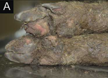
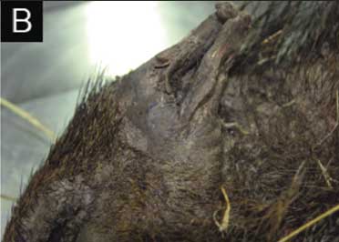
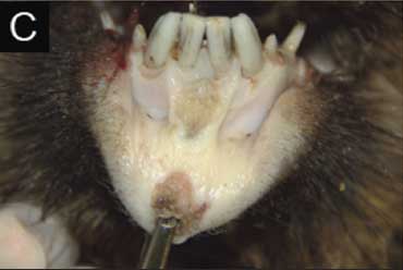
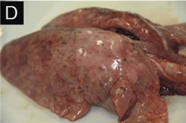
The animal was received within 10 hours after its death. The time of death was accurately estimated, as workers who had fed the animals 10 hours earlier had not found a dead pig near the feeders. The affected animal was a 6-month-old female (age estimate based on tooth replacement and eruption patterns).13 External examination revealed poor body condition, with greasy brown exudates and skin ulcers around the mouth and eyes and on the neck and legs, and separation of the horn at the bulbs of the heels. Necrosis with loss of tissue affecting the tips of the pinnae and tail were also present. Cervical and inguinal lymph nodes were moderately swollen. Lungs were diffusely hyperemic, with multifocal white 1- to 3-mm foci surrounded by hemorrhagic halos throughout the parenchyma (Figure 1).
Figure 1: Gross lesions in a wild boar (6 months of age) with exudative epidermitis. This animal was found dead in a game estate in Spain housing approximately 350 wild boar. A: Ulceration and separation of the hoof horn at the bulbs of the heels. B: Necrosis with loss of tissue at the tip of the pinna. C: Focal ulcerative stomatitis. D: Lung with generalized hyperemia and multifocal necrotic and hemorrhagic foci.
|
Pathological and microbiological examinations
Tissue samples from brain, heart, kidney, liver, lungs, lymph nodes, skin, and spleen were collected and processed for histopathological examination. Briefly, tissues were placed in 10% buffered formalin, trimmed, and embedded in paraffin, sectioned at 3 to 4 µm, and stained with hematoxylin and eosin. Skin and lung sections were also stained with Gram stain. In addition, in order to conduct a microbiological study, tissue samples from heart, kidney, liver, lung, skin, and spleen were cultured on blood agar and MacConkey agar plates and incubated aerobically for 24 hours at 37°C. Isolates obtained were identified using standard methods for phenotypic characterization as previously described.14 Identification was confirmed using the Phoenix 100 system for bacterial identification (Becton Dickinson, New Jersey).
Finally, as development of exudative epidermitis has been associated with viral co-infections, eg, PPV or PCV2,6,7 serum was tested for antibodies against these viruses. A blood sample obtained directly from the heart of the affected wild boar was centrifuged at 1500g for 5 minutes, and the harvested serum was stored at -20°C until used. Serum was tested by a commercial indirect enzyme-linked immunosorbent assay kit (ELISA), Ingezim Circovirus IgG/IgM and Ingezim PPV (Ingenasa, Madrid, Spain) according to the manufacturer’s instructions.
Histopathological results
Histopathology revealed vesiculopustular dermatitis in the damaged skin, characterized by diffuse moderate irregular acanthosis (epidermal hyperplasia) and ortho- and parakeratotic hyperkeratosis with multifocal intraepidermal pustules that contained degenerate neutrophils, necrotic debris, and microcolonies of gram-positive cocci. In the dermis, there was mild superficial perivascular to interstitial edema and mild inflammatory infiltrate, mainly with mononuclear cells and fewer neutrophils. Ulcerated areas were covered with dried necrotic crusts; the suppurative exudate and bacteria extended to follicles and to deep layers of the dermis (abscess). The histopathological study confirmed severe embolic pneumonia with numerous foci of necrosis, including degenerated neutrophils and microcolonies of gram-positive cocci. In the surrounding parenchyma, alveoli were filled with neutrophils, macrophages, and cellular debris. Numerous gram-positive cocci were detected within the cytoplasm of macrophages (Figure 2). Minimal signs of autolysis were observed in these samples.
Figure 2: Microscopic lesions in a wild boar with exudative epidermitis (described in Figure 1). A: Skin showing moderate irregular acanthosis (epidermal hyperplasia) and hyperkeratosis (hematoxylin and eosin [H&E] stain, original magnification 40×). B: Skin showing intraepidermal pustule with numerous gram-positive cocci (Gram stain; bar = 100 µm). C: Lung showing foci of necrosis resulting from septic emboli. Inset, bacteria within a vessel (H&E stain, original magnification 40×). D: Lung showing numerous gram-positive cocci within alveolar macrophages (Gram stain; bar = 10 µm). 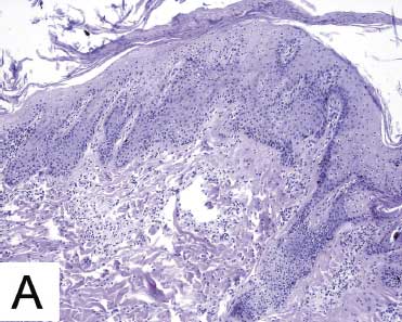 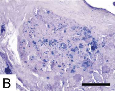 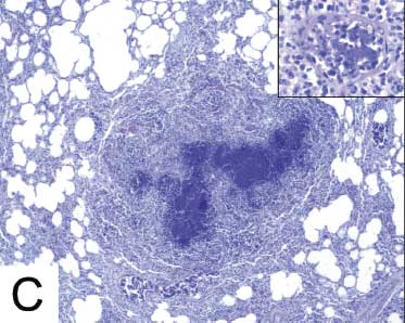 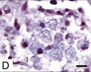 |
Microbiological and serological results
Pure growth of medium-sized, porcelain-white nonhemolytic colonies was obtained from lungs on blood agar plates. Similar colonies were predominant on blood agar cultures from skin. White nonhemolytic colonies were identified as S hyicus subspecies hyicus in both lung and skin cultures. The remainder of the isolates obtained from skin were identified as Proteus species and Pseudomonas aureginosa. Microbiological cultures from other tissues remained sterile. Antibodies against PCV2 and PPV were not detected by ELISA testing.
Discussion
In this study, we have confirmed and described a case of exudative epidermitis in a wild boar. To the authors’ knowledge, this is the first report of exudative epidermitis in a wild boar. Both macroscopic and microscopic lesions found in the studied animal were very similar to those previously described in domestic pigs. Lesions in this case were consistent with the subacute form of exudative epidermitis, characterized by skin lesions in delimited areas (eg, around eyes, snout, mouth, and heels).
Other infectious diseases, such as swine vesicular disease (SWD), may produce skin lesions similar to those found in the studied animal.15 However, SWD rarely produces skin lesions in areas such as tips of the pinnae or tail.15 In addition, the causal agent of SWD has not been detected in Spain since 1993, and hence its presence in wild boar is very unlikely.
In general, subacute forms of exudative epidermitis are seen in animals older than 35 days and in immune young animals. In this case, the animal was a 6-month-old wild boar. In addition, factors that usually predispose to more acute forms of exudative epidermitis, such as co-infection with PCV2 or PPV, were not detected by ELISA testing for antibodies to these viruses. Other factors that may also predispose swine to more severe forms of exudative epidermitis, such as high animal density or nutritional deficiencies, were not involved, since the animals were fed daily. Therefore, to our knowledge, no predisposing factors existed in this case.
Staphylococcus hyicus was isolated from the damaged skin along with P aeruginosa and Proteus species. Secondary infection with these microorganisms is common in cases of exudative epidermitis.1 The ability to produce exfoliative toxin would have had to be tested to assess the virulence of the S hyicus isolates obtained and to confirm the diagnosis.16 However, in the absence of methods to differentiate virulent from avirulent strains, all types of S hyicus should be regarded as potentially virulent.1 Furthermore, the appearance of gram-positive colonies coincident with S hyicus in the typical histological lesions of exudative epidermitis clearly suggests the implication of S hyicus in development of the skin lesions.
Piglets with the subacute forms of exudative epidermitis frequently survive, although recovery is slow and there is usually a marked depression in growth rate.8 However, in this case, exudative epidermitis was aggravated by the pneumonic lesions attributed to S hyicus. The presence of S hyicus in the lung was detected not only by culture but also histologically in the alveolar exudate, where large numbers of gram-positive cocci were observed in the cytoplasm of macrophages and in septic emboli. Staphylococcus hyicus has also been isolated from tonsils and bronchial lavage fluids of healthy pigs17 and from the pneumonic lung of a dead pig;18 however, to the authors’ knowledge, pneumonic lesions produced by this microorganism have not previously been described in an animal with exudative epidermitis.
The multiple, widely distributed lesions in the lungs, along with observation of septic emboli within vessels, suggests a hematogenous rather than an aerogenous route of infection. The abscesses in the deep dermis were most likely the source of those septic emboli. The debility produced by this chronic disease and associated immunosuppression might favor occurrence of septic emboli and hence the spread of S hyicus to the lungs.
No additional cases of exudative epidermitis were detected in the studied estate. However, we cannot be sure that no other cases had occurred. The extensive size of this game estate makes it very difficult to accurately assess mortality, since dead animals could be removed quickly by predators, remaining undetected.
Exudative epidermitis affects mainly intensive swine farms with large numbers of animals, early weaning, and high animal densities.9 In recent years, the number of intensive wild boar farms has increased notably in order to supply more animals for hunting or consumption.19,20 In addition, in these farms, factors predisposing to exudative epidermitis have been found, such as a high prevalence of PCV2 and PPV.19 Thus the risk of exudative epidermitis in wild boar may be higher than expected in these farms, which might produce serious economic losses.
This disease and others that occur on wild-boar farms, while similar to those described in domestic pigs, tend to produce specific clinical signs in wild boar, for example, ocular damage in wild boar with swine erysipelas11 or pneumonic lesions caused by S hyicus as described in this case. Thus, increasing knowledge about the epidemiological, clinical, and pathological characteristics of these diseases in wild boar will facilitate prevention, diagnosis, and treatment, reducing the health and economic impact on this new swine-production niche.
Implications
• Exudative epidermitis may occur in wild boar, with lesions similar to those found in domestic pigs.
• Wild boar affected by exudative epidermitis may have severe pneumonic lesions caused by S hyicus, suggesting a hematogenous route of infection.
Acknowledgments
This work was supported by Ministerio de Ciencia e Innovación (Gobierno de España) and Gobierno de Extremadura. Dr Risco has a Formación de Profesorado Universitario grant from the Ministerio de Ciencia e Innovación.
Conflict of interest
None reported.
References
1. Wegener HC, Skov-Jensen EW. Exudative epidermitis. In: Straw B, Zimmermann J, D’Allaire S, Taylor DJ, eds. Diseases of Swine. 9th ed. Ames, Iowa: Blackwell Publishing; 2006:675–679.
2. Pepper TA, Taylor DJ. The effect of exudative epidermitis on weaner production in a small pig herd. Vet Rec. 1977;101:204–205.
3. Aarestrup FM, Wegener HC. Association between production of fibrinolysin and virulence of Staphylococcus hyicus in relation to exudative epidermitis in pigs. Acta Vet Scand Suppl. 1997;38:295–297.
4. Andresen LO, Wegener HC, Bille-Hansen V. Staphylococcus hyicus-skin reactions in piglets caused by crude extracellular products and by partially purified exfoliative toxin. Microb Pathog. 1993;15:217–225.
5. Tanabe T, Sato H, Watanabe K, Hirano M, Hirose K, Kurokawa S, Nakano K, Saito H, Maehara N. Correlation between occurrence of exudative epidermitis and exfoliative toxin-producing ability of Staphylococcus hyicus. Vet Microbiol. 1996;48:9–17.
6. Wattrang E, McNeilly F, Allan GM, Greko C, Fossum C, Wallgren P. Exudative epidermitis and porcine circovirus-2 infection in a Swedish SPF-herd. Vet Microbiol. 2002;86:281–293.
7. Whitaker HK, Neu SM, Pace LW. Parvovirus infection in pigs with exudative skin disease. J Vet Diagn Investig. 1990;2:244–246.
8. Ginn PE, Mansell JEKL, Rakich PM. Skin and appendages. In: Maxie MG, Jubb KVF, eds. Jubb, Kennedy and Palmer’s Pathology of Domestic Animals. Edinburgh, United Kingdom: Elsevier; 2007:679–680.
9. Kim J, Chae C. Concurrent presence of porcine circovirus type 2 and porcine parvovirus in retrospective cases of exudative epidermitis in pigs. Vet J. 2004;167:104–106.
10. Wegener HC, Andresen LO, Bille-Hansen V. Staphylococcus hyicus virulence in relation to exudative epidermitis in pigs. Can J Vet Res. 1993;57:119-125.
11. Risco D, Llario PF, Velarde R, García WL, Benítez JM, García A, Bermejo F, Cortés M, Rey J, De Mendoza JH, Gómez L. Outbreak of swine erysipelas in a semi-intensive wild boar farm in Spain. Trans Emerg Dis. 2011;58:445–450.
12. García-Jiménez WL, Benítez-Medina JM, Fernández-Llario P, Abecia JA, García-Sánchez A, Martínez R, Risco D, Ortiz-Peláez A, Salguero FJ, Smith NH, Gómez L, Hermoso de Mendoza J. Comparative pathology of the natural infections by Mycobacterium bovis and by Mycobacterium caprae in wild boar (Sus scrofa). Trans Emerg Dis. 2012;60:102–109.
13. Boitani L, Mattei L. Aging wild boar (Sus scrofa) by tooth eruption. In: Spitz F, Janeau G, González G, Aulagnier S, eds. Ongules/Ungulates. 9th ed. Tolouse, France: SFEPM-IRGM; 1992:419–421.
14. Barrow GI, Feltham RKA. Cowan and Steel’s Manual for the Identification of Medical Bacteria. 3rd ed. Cambridge, United Kingdom: Cambridge University Press; 1993:52–57.
15. Lubroth J, Rodríguez L, Dekker A. Vesicular diseases. In: Straw B, Zimmerman J, D´Allaire S, Taylor DJ, eds. Diseases of Swine. 9th ed. Ames, Iowa: Blackwell Publishing; 2006:517–536.
16. Andresen LO, Ahrens P. A multiplex PCR for detection of genes encoding exfoliative toxins from Staphylococcus hyicus. J Appl Microbiol. 2004;96:1265–1270.
17. Hensel A, Ganter M, Kipper S, Krehon S, Wittenbrink MM, Petzoldt K. Prevalence of aerobic bacteria in bronchoalveolar lavage fluids from healthy pigs. Am J Vet Res. 1994;55:1697–1702.
18. Skalka B. Výskyt stafylokokových druhu u klinicky zdravých hospodárských zvírat [Occurrence of staphylococcal species in clinically healthy domestic animals]. Veterinarni Medicina [Veterinary Medicine – Praha]. 1991;36:9–19.
19. Halli O, Ala-Kurikka E, Nokireki T, Skrzypczak T, Raunio-Saarnisto M, Peltoniemi OAT, Heinonen M. Prevalence of and risk factors associated with viral and bacterial pathogens in farmed European wild boar. Vet J. 2012;194:98–101.
20. Gortázar C, Acevedo P, Ruiz-Fons F, Vicente J. Disease risks and overabundance of game species. Eur J Wildl Res. 2006;52:81–87.