Huang Y, Henry S, Friendship R, et al. Clinical presentation, case definition, and diagnostic guidelines for porcine periweaning failure to thrive syndrome. J Swine Health Prod. 2011;19(6):340–344
| Diagnostic notes | Peer reviewed |
SummaryPorcine periweaning failure to thrive syndrome (PFTS) is a clinical condition characterized by anorexia, lethargy, and progressive debilitation of pigs occurring within 2 to 3 weeks after weaning. In affected populations, there is a striking contrast between the clinically affected pigs, which progress from being normally active to lethargic within days of weaning, and the unaffected members of their cohort, which grow and behave normally. The etiology, pathophysiology, and pathogenesis of PFTS have not been determined, although several infectious agents have been identified in affected pigs. Histopathologic lesions of chronic active rhinitis, superficial gastritis, atrophic enteritis, superficial colitis, and thymic atrophy are observed in most PFTS-affected pigs. The basis for a presumptive diagnosis of PFTS includes the age of onset, the presence of typical clinical signs, the presence of the collective histopathologic lesions, and, importantly, the ruling out of other known swine diseases (for example, porcine circovirus associated disease, swine influenza, porcine reproductive and respiratory syndrome, and bacterial infections). The objectives of this paper are to propose a clinical case definition, describe the characteristic clinical progression, signs, and observed lesions of PFTS, and to make recommendations for investigation of PFTS-suspected farms. | ResumenEl síndrome porcino de retraso en el desarrollo en el peridestete (PFTS por sus siglas en inglés) es una condición clínica caracterizada por anorexia, letargo, y debilitación progresiva de cerdos dentro de las 2 a 3 semanas después del destete. En poblaciones afectadas, existe un gran contraste entre los cerdos afectados clínicamente, que evolucionan de estar normalmente activos a letárgicos a los pocos días después del destete, en contraste con los cerdos no afectados de su grupo, que crecen y se comportan normalmente. La etiología, patofisiología, y patogénesis del PFTS no han sido determinadas, sin embargo, se han identificado varios agentes infecciosos en cerdos afectados. Se observaron lesiones histopatológicas de rinitis activa crónica, gastritis superficial, enteritis atrófica, colitis superficial, y atrofia del timo en la mayoría de los cerdos afectados por el PFTS. La base para un diagnóstico presuntivo del PFTS incluye la edad de presentación, la presencia de signos clínicos típicos, la presencia de lesiones histopatológicas múltiples compatibles, y lo que es más importante, el descartar otras enfermedades porcinas conocidas (por ejemplo, enfermedades asociadas a cirvovirus porcino, influenza porcina, síndrome reproductivo y respiratorio porcino, e infecciones bacterianas). Los objetivos de este estudio son proponer una definición clínica del caso, describir la evolución clínica característica, signos y lesiones observadas del PFTS, y hacer recomendaciones para la investigación en granjas con sospecha del PFTS. | ResuméLe syndrome de déficience de croissance péri-sevrage (PFTS) est une condition clinique caractérisée par de l’anorexie, de la léthargie, et un dépérissement progressif des porcs se produisant lors des 2 à 3 semaines suivant le sevrage. Dans les populations affectées, il y a un contraste frappant entre les porcs cliniquement affectés qui, en quelques jours, passent d’un état normal à être léthargiques, et les membres non-affectés de leur cohorte, qui ont un comportement et une croissance normale. L’étiologie, la patho-physiologie, et la pathogénèse du PFTS n’ont pas été déterminées, bien que plusieurs agents infectieux aient été identifiés chez les porcs atteints. Des lésions histopathologiques de rhinite active chronique, de gastrite superficielle, d’entérite atrophique, de colite superficielle, et d’atrophie du thymus sont observées chez la majorité des porcs affectés par le PFTS. Le fondement pour un diagnostic présomptif de PFTS inclus l’âge du début de la condition, la présence de signes cliniques typiques, la présence de l’ensemble des lésions histopathologiques, et, de manière importante, l’élimination des autres maladies porcines connues (par exemple, la maladie associée au circovirus porcin, l’influenza porcin, le syndrome reproducteur et respiratoire porcin, et les infections bactériennes). Les objectifs de cet article sont de proposer une définition d’un cas clinique, décrire la progression clinique, les signes et les lésions caractéristiques observés lors de PFTS, et de faire des recommandations pour enquêter sur les fermes suspectées d’être au prise avec le PFTS. |
Keywords: swine, porcine periweaning failure to thrive syndrome, weight loss, nursery, postweaning
Search the AASV web site
for pages with similar keywords.
Received: October 7, 2010
Accepted: May 2, 2011
Porcine periweaning failure to thrive syndrome (PFTS) presents clinically as moderate but variable morbidity (range 1% to 20%) with high case fatality. Individual pigs affected within 7 days after weaning demonstrate anorexia, lethargy, and progressive debilitation. PFTS has specifically been noted in proceedings and publications from veterinarians and researchers since 2008,1-3 although clinicians have observed weanling pigs with similar clinical signs for many years. In some farms, the clinical signs of PFTS may have been confused with or masked by other diseases, including porcine circovirus associated disease, swine influenza, and porcine reproductive and respiratory syndrome (PRRS). Recognition shortly after weaning of cachectic and debilitated pigs that were negative for PRRS virus (PRRSV) and swine influenza virus and immunized against porcine circovirus type 2 suggests that PFTS is a distinct clinical entity. The authors are aware of PFTS or clinically similar cases reported in a number of different regions of North America. Little is known about the cause of PFTS, but infectious agents and management-related factors need to be ruled out. In this paper, we propose a clinical case definition for PFTS, describe the characteristic clinical progression and signs and observed lesions, and make recommendations for investigation of PFTS-suspected farms.
Obtaining the consensus name “porcine periweaning failure to thrive syndrome (PFTS)”
PFTS was previously reported as postweaning catabolic syndrome,1 postweaning wasting-catabolic syndrome,4,5 and failure to thrive syndrome,3 and is possibly the same disease as postweaning fading pig-anorexia syndrome.6 At the 2010 International Pig Veterinary Society Congress in Vancouver, a number of researchers, diagnosticians, and practitioners from North America met and reached consensus that the name “periweaning failure to thrive syndrome (PFTS)” reflects the age of onset and clinical presentation of the syndrome. The authors encourage the adoption of this name when describing cases meeting the case definition outlined below, until such time that new knowledge of the etiology and pathogenesis supports a more specific disease designation. We suggest that “periweaning” is preferable to “postweaning,” because there may be management or infectious factors both preweaning and post weaning that contribute to the development or risk of PFTS.
Revised case definition
A proposed case definition for PFTS, revised and rephrased from Friendship et al,5 is as follows: “PFTS is characterized clinically by the progressive debilitation of weanling (nursery) pigs in the absence of discernible and detrimental infectious, nutritional, managemental, or environmental factors that can explain the clinical syndrome. At weaning, affected pigs are of average to above average body weight, and neither affected pigs nor their cohorts show evidence of residual illnesses from the suckling phase. Within 7 days of weaning, affected pigs are anorexic and lethargic. They deteriorate and within 2 to 3 weeks of weaning demonstrate marked muscle weakness and loss of body condition. Some affected pigs in all affected farms show repetitive oral behavior such as licking, chewing, or chomping. In affected farms, morbidity and mortality by batch varies over time, but case fatality is high.”
Clinical signs
The most frequently observed clinical signs of PFTS are listed in Table 1. It is important to emphasize that affected pigs and their cohorts demonstrate no obvious detrimental clinical disease at the herd level prior to weaning. At weaning, the pigs that will become affected appear healthy and of average to above average body weight and condition, and cannot be identified as “at risk.” Four to 5 days after weaning, the affected pigs have hollow abdomens or flanks, presumably the result of anorexia. Affected pigs appear hydrated. Frequent sneezing may be heard in the nursery, but coughing and dyspnea are not typically observed. In affected pigs, abnormal oral behavior of repetitive licking or chewing motions, or a repetitive and intentional “chomping” activity, during which their heads may be drooped or elevated by resting the jaw on the back of a penmate, may be observed, provided the observer remains motionless and quiet for a period of time. Such pigs are easily disturbed, making observation difficult. While identifying PFTS-affected pigs within the first week after weaning requires careful observation, affected pigs are easily identified by their lethargy, hollow abdomens, and failure to grow (Figure 1) by the second week post weaning. With quiet observation, affected pigs may be observed grouped together in a side-by-side stance with heads drooped. While this unusual posture may be maintained for long periods of time, pigs eventually become weak, lie down together, and may pile as if chilled. By 3 weeks post weaning, most of the affected pigs are severely debilitated (Figure 1), have died, or have been euthanized.
Figure 1: Periweaning failure to thrive syndrome in nursery pigs. A: an affected pig showing a hollow abdomen; B: a severely debilitated pig. 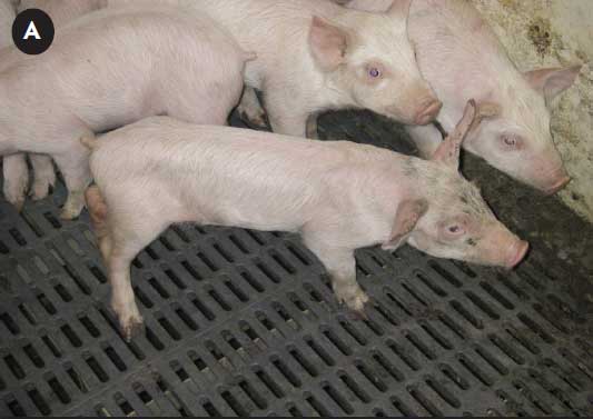 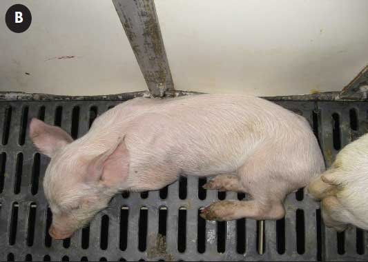 |
Table 1: Most frequently observed clinical signs and pathological changes observed in periweaning failure to thrive syndrome
* Lesions that have also been observed in age-matched cohorts. |
Most management interventions, environmental manipulations, and medical treatments are ineffective. That said, a critical review of all production practices must be performed to reduce stress, to prevent exacerbation of all nursery diseases including PFTS, and to ensure animal welfare is maintained. Techniques that are consistently effective in reducing PFTS morbidity and mortality have not been accomplished.
Relevant gross and histological observations
The definition of PFTS remains currently at the clinical level, but the frequently observed gross and histological changes found in PFTS-affected pigs are summarized in Table 1. Whether or not these lesions are significant to the pathogenesis of PFTS needs to be determined. Necropsy examination reveals scant ingesta within the gastrointestinal tract. The small intestines of affected pigs are empty or sometimes fluid-filled. The colon is empty or may contain pasty to liquid content. At later stages in the disease process, when most sick pigs are submitted for diagnostics, affected pigs have marked thymic atrophy, which is most obvious in the thorax. If affected pigs in earlier stages are examined, thymic atrophy appears to be less severe grossly, suggesting that it may be associated with prolonged anorexia or sickness, rather than a primary lesion. Bronchopneumonia may be observed in some pigs, especially those in later disease stages, but is not a consistent finding. When the head is sectioned on the midline, suppurative rhinitis, characterized by purulent material in the nasal cavity as proximal as the ethmoid conchae, is frequently observed in affected pigs as well as in some healthy penmates.
Microscopic lesions noted in PFTS-affected pigs include lymphocytic and suppurative rhinitis with or without cytomegalovirus inclusion bodies, lymphocytic superficial fundic gastritis, atrophic enteritis, and superficial colitis. Although the reasons for and the significance of these particular lesions are not clear at this time, they are prevalent in affected pigs and serve as pathologic indicators of PFTS. It is noteworthy however, that lymphocytic and suppurative rhinitis with or without cytomegalovirus inclusion bodies and superficial colitis have also been observed in healthy age-matched penmates of the affected pigs.7
Recommendations for herd investigations
As the etiology and pathogenesis of PFTS are unknown, the presence of typical clinical progression, the age of onset, and the elimination by thorough diagnostic investigation of all other known porcine diseases as primary entities should be used to classify affected farms as having PFTS. A non-exclusive list of potential differential diagnoses based on clinical signs and pathological changes are listed in Table 2.
Table 2: Potential differential diagnoses based on clinical signs and pathological changes associated with pigs affected with periweaning failure to thrive syndrome*
* This is not intended to be a complete list of differential diagnoses. Swine diseases and pathogens exotic to Canada and the United States are excluded. PCV2 = porcine circovirus type 2; PRRSV = porcine reproductive and respiratory syndrome virus; HEV = hemagglutinating encephalomyelitis virus; PCMV = porcine cytomegalovirus; TGEV = transmissible gastroenteritis virus.
|
Recognizing PFTS-affected pigs in the early stage (ie, 4 to 5 days post weaning) is difficult but is vital to the proper interpretation of uncomplicated gross and microscopic pathology. Pigs that are sick for a prolonged period are at greater risk of succumbing to secondary infections that mask the original pathology. In our experience, an encompassing list of tissues must be evaluated microscopically (Table 3), even if they look grossly normal. Failing to collect certain tissues in suspected cases prevents effective comparison with other cases, which is critical when characterizing a new syndrome such as PFTS. It is important to examine nasal turbinate, fundic stomach, small intestine, large intestine, and thymus histologically, but these should not be the only organs examined. In addition, examining age-matched healthy cohorts from suspect farms is necessary to correctly interpret the significance of some gross and histologic lesions observed in sick pigs. For example, if specific lesions are of equivocal prevalence and severity in both sick pigs and healthy cohorts, the significance of these lesions is less certain. Special arrangements with the diagnostic laboratory are strongly recommended to reduce the substantial diagnostic costs associated with examination of an extensive tissue set and the number of animals required in a disease investigation compared to routine diagnostic submissions. In our investigations of PFTS, it has also been useful to examine healthy pigs from unaffected farms in order to characterize the morphology of normal, healthy age-matched pigs. Recommendations for investigation of herds demonstrating signs typical of PFTS are listed in the text box, which can serve as a checklist of actions to perform during investigation.
Table 3: Tissues to collect from pigs suspected of having periweaning failure to thrive syndrome*
* The tissues listed in this table are not exhaustive. Other tissues may be collected if deemed to provide useful information. † The differential diagnoses in this table are examples and are by no means complete. PRRSV = porcine reproductive and respiratory syndrome virus; SIV = swine influenza virus; PCV2 = porcine circovirus type 2; |
||||||||||||||||||||||||||||||||||||||
Implications
• Porcine PFTS is characterized by anorexia and lethargy of nursery pigs beginning soon after weaning and is not associated with porcine circovirus type 2, PRRSV, or SIV infection.
• The etiology and pathogenesis are unknown.
• The age of disease onset, presence of characteristic clinical signs, and elimination by thorough diagnostic investigation of other known porcine diseases as primary entities should be used to identify farms affected by PFTS.
• A thorough history, clinical examination, and gross and microscopic evaluation of affected pigs are necessary to work up suspected cases of PFTS, with pathologic examination targeting affected pigs and age-matched healthy cohorts between weaning and 3 weeks post weaning.
• Continued collaboration among clinicians, producers, and diagnosticians is needed to identify cause(s), risk factors, and prevention strategies for this syndrome.
Acknowledgements
The authors sincerely thank all the producers, veterinarians, diagnosticians, and technicians who have supported the numerous farm and pathological investigations that have been undertaken to date. The leadership of Drs Jerome Nietfeld, Dick Hesse, Bob Rowland, and Cino Ozuna in Kansas, Drs Brendan O’Connor, Beverley Kidney, Mark Swendrowski, Henry Gauvreau, Peter Provis, and Michael Sheridan in Western Canada, and Drs Josepha Delay and Brent Jones in Ontario is noteworthy. Funding for this research has been generously provided by the Western College of Veterinary Medicine Disease Investigation Fund, the Saskatchewan Pork Development Board, and Saskatchewan Agriculture and Food; and through in-kind contributions from The Ontario Animal Health Laboratory, Manitoba Veterinary Laboratory Services, Alberta Agriculture and Rural Development, Kansas Veterinary Diagnostic Laboratory, and Kansas Swine Diagnostic Fund, and contributions of many producers with affected herds.
References
*1. Dufresne L, Fangman TJ, Henry S. Post-weaning catabolic syndrome: complexities and perspectives. Proc Allen D. Leman Swine Conf. St Paul, Minnesota. 2008:79–85.
*2. Gauvreau H, Harding J. Why are these nursery pigs dying? An ongoing field investigation into a farm with elevated nursery mortality associated with non-PRRS/PCV2 post weaning starvation. Proc Western Canadian Assoc Swine Vet Conf. Saskatoon, Saskatchewan. 2008.
*3. Vansickle J. Researchers scramble to solve failure to thrive syndrome. National Hog Farmer. Sept 15, 2008. Available at: http://nationalhogfarmer.com/health-diseases/0915-researchers-trying-solve-syndrome/. Accessed 15 June 2011.
*4. Harding JCS. Swine health challenges and emerging diseases in Western Canada: what’s on the horizon? Proc Saskatchewan Pork Industry Symp. Saskatoon, Saskatchewan. 2010:19–20.
*5. Friendship B, Harding J, Henry S. Periweaning failure to thrive syndrome (PFTS) – difficulties of investigating an emerging clinical problem. Proc Allen D. Leman Swine Conf. St Paul, Minnesota. 2010:73–78.
*6. Rossow K. Postweaning “fading pig/anorexia syndrome.” National Hog Farmer. June 28, 2010. Available at: http://nationalhogfarmer.com/weekly-preview/0628-postweaning-fading-pig-anorexia/. Accessed 15 June 2011.
7. Huang Y, Gauvreau H, Harding J. Diagnostic investigation of porcine periweaning failure to thrive syndrome (PFTS): Lack of compelling evidence linking to common porcine pathogens. J Vet Diag Invest. In press.
8. Pensaert MB. Hemagglutinating encephalomyelitis virus. In: Straw BE, Zimmerman JJ, D’Allaire SD, Taylor DJ, eds. Diseases of Swine. 9th ed. Ames, Iowa: Blackwell Publishing Professional; 2006:353–358.
* Non-refereed references.