Gauger PC, Patterson AR, Kim WI, et al. An outbreak of porcine malignant catarrhal fever in a farrow-to-finish swine farm in the United States. J Swine Health Prod. 2010;18(5):244–248
| Case report | Peer reviewed |
Cite as: Gauger PC, Patterson AR, Kim WI, et al. An outbreak of porcine malignant catarrhal fever in a farrow-to-finish swine farm in the United States. J Swine Health Prod. 2010;18(5):244–248.
Also available as a PDF.
SummaryMalignant catarrhal fever is a sporadic, often fatal viral disease affecting multiple species, including swine. Ovine herpesvirus type 2 (OvHV-2), the cause of sheep-associated malignant catarrhal fever, incites nonspecific clinical signs and occasional death in swine. An outbreak of malignant catarrhal fever in a farrow-to-finish swine farm in the United States was confirmed by identifying OvHV-2 DNA in two clinically affected adult swine previously exposed to sheep. Forty-one swine exhibited clinical signs of lethargy, anorexia, and fever, with recovery or death in 22 and 19 animals, respectively. Abortion was also reported in two clinically affected pregnant females. Ovine herpesvirus type 2 DNA was identified by polymerase chain reaction (PCR) in spleen, brain, and lung tissue. A BLAST homology search of the sequenced PCR amplicon matched the conserved region of the OvHV-2 tegument protein. Porcine malignant catarrhal fever is difficult to diagnose due to the nonspecific clinical signs, rarity of occurrence, and sporadic nature of the disease. Polymerase chain reaction assays and serologic testing are available to assist in an accurate diagnosis. Veterinarians should consider malignant catarrhal fever a potential differential diagnosis in swine with poorly defined clinical signs, intermittent death, and previous exposure to sheep. | ResumenLa fiebre catarral maligna es una enfermedad viral esporádica, a menudo fatal que afecta a múltiples especies, incluyendo la porcina. El herpesvirus ovino tipo 2 (OvHV-2), la causa de la fiebre catarral maligna asociada con ovejas, provoca signos clínicos no específicos y muerte ocasional en cerdos. Se confirmó un brote de fiebre catarral maligna en una granja porcina de ciclo completo en los Estados Unidos al identificar el OvHV-2 DNA en dos cerdos adultos afectados clínicamente, previamente expuestos a ovejas. Cuarenta y un cerdos exhibieron signos clínicos de letargo, anorexia, y fiebre, con recuperación ó muerte en 22 y 19 animales, respectivamente. También se reportaron abortos en dos hembras gestantes afectadas clínicamente. El DNA del herpesvirus ovino tipo 2 se identificó a través de la reacción en cadena de la polimerasa (PCR por sus siglas en inglés) en tejidos de bazo, cerebro, y pulmón. Una búsqueda homóloga en BLAST del amplicón secuenciado en PCR coincidió con el área conservada de la proteína de tegumento OvHV-2 tegument. La fiebre catarral maligna porcina es difícil de diagnosticar debido a los signos clínicos no específicos, lo raro de la ocurrencia, y la naturaleza esporádica de la enfermedad. La prueba de reacción en cadena de la polimerasa y las pruebas serológicas están disponibles para ayudar a obtener un diagnóstico preciso. Los veterinarios deberían considerar a la fiebre catarral maligna como un diagnóstico diferencial potencial en cerdos con signos clínicos poco definidos, muerte intermitente, y previa exposición a ovejas. | ResuméLa fièvre catarrhale maligne est une maladie virale sporadique, souvent fatale, pouvant affecter plusieurs espèces, incluant le porc. L’herpèsvirus ovin de type 2 (OvHV-2), la cause de la fièvre catarrhale maligne associée au mouton, entraîne des signes cliniques non-spécifiques et des mortalités occasionnelles chez les porcs. Une épidémie de fièvre catarrhale maligne dans une ferme porcine de type naisseur-finisseur située aux États-Unis a été confirmée par identification d’ADN du OvHV-2 chez deux animaux adultes présentant des signes cliniques préalablement exposés à des moutons. Quarante-et-un porcs ont présenté des signes de léthargie, anorexie, et fièvre, suivi d’une guérison ou de mort chez 22 et 19 animaux, respectivement. Des avortements ont également été rapportés chez deux femelles gestantes affectées cliniquement. L’ADN du OvHV-2 a été identifié par réaction d’amplification en chaîne par la polymérase (PCR) dans la rate, le cerveau, et les poumons. Une recherche d’homologie BLAST de l’amplicon séquencé obtenu par PCR concordait avec la région conservée de la protéine du tégument d’OvHV-2. La fièvre catarrhale maligne porcine est difficile à diagnostiquer étant donné les signes cliniques non-spécifiques, sa rareté, et la nature sporadique de la maladie. Les épreuves PCR et les tests sérologiques sont disponibles pour aider au diagnostic précis. Les vétérinaires devraient considérer la fièvre catarrhale maligne comme un diagnostic différentiel possible chez les porcs présentant des signes cliniques peu définis, des mortalités intermittentes, et une exposition préalable à des moutons. |
Keywords: swine, malignant catarrhal fever, ovine
herpesvirus type 2, outbreak, polymerase chain reaction, PCR
Search the AASV web site
for pages with similar keywords.
Received: February 2, 2010
Accepted: April 1, 2010
Malignant catarrhal fever (MCF) is a systemic, often fatal lymphoproliferative disease of the ruminant families Bovidae and Cervidae.1 It is caused by a group of gamma herpesviruses within the genus Rhadinovirus. Two endemic forms of MCF are recognized and include a wildebeest-associated form caused by alcelaphine herpesvirus type 1 (AlHV-1) and a sheep-associated form caused by the ovine herpesvirus type 2 (OvHV-2).2 Both forms cause inapparent infection in their reservoir hosts, which include wildebeests and sheep, respectively. However, in ruminants other than the reservoir hosts, the two forms of MCF are clinically and pathologically indistinguishable.1,3 Sheep-associated MCF has been reported worldwide,3 and disease commonly occurs in ungulates, including cattle, bison, and deer.4,5
A case of porcine MCF attributed to OvHV-2 was first confirmed in Norway in 1998.6 Since that time, subsequent cases have been reported in Switzerland7 and Finland.8 Although unconfirmed, cases suggestive of porcine MCF were reported earlier in Germany,9 Norway,10 Sweden,11 and Switzerland.12 Clinical signs in swine, as reported from these previous cases, are similar to those described for cattle and include various combinations of fever, anorexia, ptyalism, diarrhea, lacrimation, corneal opacity, central nervous system (CNS) disease, generalized lymphadenopathy, and often a mucopurulent ocular and nasal discharge.1,6,7,13 In many cases, contact with sheep prior to the onset of clinical disease was reported.6
Recently, the first two cases of sheep-associated MCF were diagnosed in adult swine in the United States.13 These, however, were sporadic cases involving individual swine that originated on small farms. This is, to the authors’ knowledge, the first reported outbreak of MCF in a farrow-to-finish swine farm, associated with abortions, and located in the central United States.
Case description
The case farm consisted of 135 adult swine located on a farrow-to-finish, specific-pathogen-free purebred seedstock operation with Chester White, Duroc, Yorkshire, and Berkshire breeds. The 4.05-hectare farm included a 12.2 × 24.4-m double-curtain gestation barn (G-barn) with 10 pens on each side of a 1.2-m center alley. Each of the 2.4 × 5.5-m pens had steel vertical bars approximately 1.1 m high that bordered the center alley. Individual gestation crates were not used. The premise included a 250-head nursery and two finishing facilities: a double-curtain barn containing 400 animals and an open-front pole barn containing 300 animals. Additional livestock present at the time of the outbreak included a small flock of 43 sheep consisting of ewes, lambs, and one ram, various numbers of rabbits, ducks, and geese, and one cat.
In early January 2008, the owner placed nine ewe lambs approximately 10 to 12 months of age in the G-barn alleyway due to inclement weather and to reduce feed competition with older ewes. The ewe lambs were supplied their own water source, maintained on a hay and grain diet, and were fed at one end of the G-barn alley. Gated pens with vertical steel bars allowed ample nose-to-nose contact between the sheep and 120 adult swine housed in the G-barn, which consisted of 35 gilts and approximately 85 multiparous sows and boars penned according to age and sex. The sheep were removed from the G-barn the first week of March and the alley was cleaned but not disinfected.
Clinical signs began March 20, 2008. Forty-one adult swine became clinically affected over the next 6 months. Clinical signs included pyrexia, anorexia, and lethargy. Fevers ranged from 39°C to 40°C and the herd veterinarian reported that affected animals were polydipsic. Clinical signs typically developed 24 to 48 hours before death, although 22 animals recovered. Individual animals were nonresponsive to treatment with injectable antibiotics, and an insignificant response resulted from two subsequent mass treatments with feed-grade antibiotics. Sporadic death loss extending over 6 months began with a parity-10 sow and a boar. Mortality and morbidity that occurred over the next 5 months are shown in Table 1. Overall, 41 of 120 exposed adult swine (34%) were clinically affected. Nineteen of 120 adult swine (16%), five of which were gilts, died, and an additional 22 (18%) that displayed clinical signs apparently recovered. The case fatality rate was 19 of 41 affected swine (46%).
Table 1: Monthly mortality and morbidity for swine infected with ovine herpesvirus type 2 in the gestation barn of a farrow-to-finish swine herd*
* Swine were exposed to sheep from early January until the first week of March. † Number of affected adult swine among 120 total animals in the gestation barn. |
The herd veterinarian began a diagnostic investigation on April 24, 2008, with a serological profile. Six adult female swine were selected for testing by ELISA for antibodies to porcine reproductive and respiratory syndrome virus (PRRSV) and swine influenza virus nucleoprotein (SIV-NP) ELISA. Anti-PRRSV antibody was not detected, but anti-SIV-NP antibody was detected in a 4-year-old female. PRRS virus RNA was not detected by polymerase chain reaction (PCR) in a pooled sample of serum from the three oldest females.
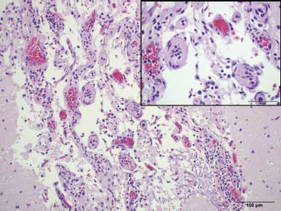
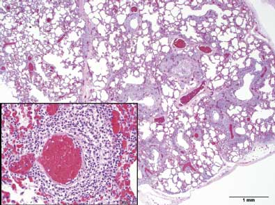
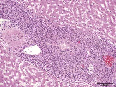
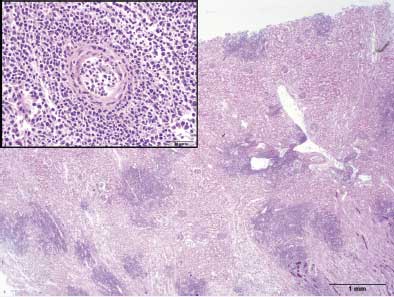
Porcine submissions to the Iowa State University Veterinary Diagnostic Laboratory (ISU-VDL) included three dead animals, three tissue-sample submissions, 15 aborted fetuses, 39 serum samples, and five nasal swabs between April and August, 2008. Clinical disease was described as anorexia, lethargy, and sudden death. All 41 affected animals were housed in the G-barn; swine in the nursery and finisher barns were unaffected. Gross necropsy findings were nonspecific and included diffuse pulmonary congestion with interlobular edema. Polymerase chain reaction assays were negative for PRRSV, SIV, and porcine circovirus type 2 (PCV2). Aerobic and anaerobic bacterial cultures, which included enrichment for Erysipelothrix rhusiopathiae, were negative. Additional tests for Leptospira interrogans serovars, bovine virus diarrhea virus, novel porcine pestivirus-like virus (agent X),14 classical swine fever, and pseudorabies virus were negative. Histopathologic lesions were similar in all tissue samples and included marked perivascular inflammation and mononuclear vasculitis in multiple tissues including the brain (Figure 1), lung (Figure 2), liver (Figure 3), kidney (Figure 4), heart, lymph node, and uterus. Rarely, affected blood vessels were necrotic with hyalinized vascular walls (Figure 1).
|
|
||
|
|
Sporadic abortions began in May, approximately 4 months post initial exposure to sheep, at 104 to 111 days of gestation and 1 to 2 days after the onset of clinical signs. Routine diagnostic testing for reproductive diseases caused by PRRSV, PCV2, and Leptospira interrogans serovars were negative by PCR or immunohistochemistry, without specific testing for OvHV-2. A final diagnostic submission of a 16-month-old female swine on August 25, 2008, revealed no gross lesions. Histopathology revealed lymphocytic vasculitis in multiple tissues, consistent with a lymphoproliferative disease. Repeated identification of mononuclear vasculitis in the submitted swine tissues prompted submission of spleen to Colorado State University for OvHV-2 PCR. A positive result confirmed a diagnosis of sheep-associated MCF. Archived brain and lung tissue from one animal were tested for OvHV-2 by a previously published real-time PCR developed at the ISU-VDL and based on the tegument protein gene of the OvHV-2 genome.15 Moreover, positive tissues were tested by a conventional semi-nested OvHV-2 PCR as previously described,15 and the 231-bp amplified products were subjected to sequencing. BLAST homology search revealed that the 231-bp amplicon matched the tegument protein gene of OvHV-2 in Genbank (GenBank accession no. S64565) (Figure 5).
Figure 5: Nucleotide comparison between a sequenced polymerase chain reaction (PCR) amplicon from a conventional semi-nested ovine herpesvirus type 2 PCR15 performed on a swine brain homogenate and a sequence of the 140 kda tegument protein gene of ovine herpesvirus type 2 available in GenBank (accession no. S64565).
|
Discussion
Although outbreaks of MCF have been described on sow farms in Europe,8 to the authors’ knowledge, this is the first report of an outbreak of sheep-associated MCF in a farrow-to-finish swine operation in the United States. Two individual and sporadic cases of sheep-associated MCF in swine were previously described in the United States.13 These cases occurred on separate nontraditional swine farms in New York and Kentucky. The New York pig was located on an animal rescue farm, and the Kentucky case involved a pregnant sow housed in a high-school agricultural facility. Affected animals in both cases were either located on the same farm with, or housed with, two additional pigs and three adult sheep in separate pens that were allowed nose-to-nose contact for an unknown length of time. The additional two pigs on the Kentucky farm presented with similar clinical signs but recovered.
The clinical signs and pathologic lesions in this case were similar to those described in previous reports of MCF in pigs.6-8,13 Lethargy, anorexia, and elevated body temperatures for approximately 2 days duration were consistent clinical features prior to death. Neurological signs and corneal opacity have been previously reported in pigs with MCF, but were not recognized in this outbreak despite the presence of histopathologic lesions in the brain.13 Only adult swine exposed to sheep were affected, and there was no predilection for age, parity, pregnancy status, or sex. Macroscopic lesions identified during post mortem examination of three swine submitted to the ISU-VDL were subtle, nonspecific, and consisted of pulmonary congestion and interlobular edema. Microscopic lesions were consistent with those of MCF and consisted of mononuclear vasculitis in acute cases and a multisystemic lymphoproliferative disease in subacute to chronic cases.
The pathogenesis of MCF in pigs has not been elucidated, although contact with sheep has been consistently documented as a precursor to clinical signs and infection.6-8,13 Sheep are the natural reservoir of OvHV-2 and remain unaffected after natural infection. Nasal shedding commonly occurs in adolescent sheep 6 to 10 months of age;16 however, adult sheep may intermittently shed large quantities of virus from nasal secretions.16 The length and type of exposure, incubation period, and infectious dose of virus in swine are unknown. Incubation periods in cattle post experimental inoculation have ranged from 2 to 12 weeks.3 In the case described in this report, clinical signs and sporadic death began approximately 8 weeks after the beginning of a 2-month period of exposure to ewe lambs and continued for approximately 7 months. However, it is unknown if the virus was transmitted via sheep nasal secretions or fecal material.
Malignant catarrhal fever infection in swine is usually sporadic, and there have been few reports of outbreaks.6,8 Previous OvHV-2 infections in swine may have gone undiagnosed due to the lack of specific clinical signs and gross lesions associated with MCF. The previous lack of available serological and molecular diagnostic tests may have also resulted in under-reporting of this disease. In contrast, previous reports6,13 have commented that the absence of porcine cases may be due to differences in production systems that prevent transmission of the virus or minimize exposure to sheep. Interestingly, a recent report detected the presence of infected but asymptomatic swine even in the absence of known exposure to sheep or goats.17 However, most cases of OvHV-2 in swine have included previous exposure to sheep, indicating their potential role in the pathogenesis of MCF in swine. In this case, sheep from the case farm were unavailable for testing by OvHV-2 ELISA or PCR.
Diagnosing MCF is challenging due to the nonspecific nature of the clinical signs. Available antemortem serological tests for OvHV-2 include virus neutralization, peroxidase-linked antibody, and competitive-inhibition ELISA.18 Unfortunately, the virus neutralization test has been validated only for AlHV-1, which can be consistently propagated in cell culture, unlike OvHV-2. The peroxidase-linked antibody test is a nonspecific test which detects antibody to the Herpesviridae family. The competitive-inhibition ELISA is the most useful serological test, as it specifically detects anti-MCF antibody in swine;18 however, the presence of antibody confirms exposure to the virus and is not diagnostic for disease. Polymerase chain reaction is a sensitive and useful test for detecting OvHV-2 DNA in whole blood and tissue samples.19 Malignant catarrhal fever in swine can be diagnosed by identifying histopathological lesions consistent with mononuclear vasculitis or lymphoproliferative disease in combination with detection of OvHV-2 DNA by PCR in affected animals.
The current report describes an outbreak of MCF in a farrow-to-finish swine operation involving exposure to sheep and subsequent clinical signs and death in multiple adult swine. Due to the lack of MCF vaccines, exposure between sheep and pigs should be avoided to prevent transmission of the disease. Malignant catarrhal fever should remain a differential diagnosis in swine exposed to sheep and clinically exhibiting lethargy, anorexia, and fever.
Implications
- Swine can become infected with OvHV-2, a herpesvirus endemic in sheep that causes inapparent infection in the reservoir host.
- Previous exposure to sheep is a consistent feature of the epidemiology associated with MCF in swine.
- Clinical signs in swine are nonspecific and consist of fever, anorexia, lethargy, and abortion.
- OvHV-2 infection can be diagnosed in swine by PCR testing of appropriate tissue samples (spleen, brain, or lung).
References
1. Plowright W. Malignant catarrhal fever virus. In: Dinter Z, Morein B, eds. Virus Infections of Ruminants. 1st ed. New York, New York: Elsevier Science Publishers BV; 1990:123–150.
2. Reid HW, Buxton D, Berrie E, Pow I, Finlayson J. Malignant catarrhal fever. Vet Rec. 1984;114:581–583.
3. Russell GC, Stewart JP, Haig DM. Malignant catarrhal fever: a review. Vet J. 2009;179:324–335.
4. Brown CC, Bloss LL. An epizootic of malignant catarrhal fever in a large captive herd of white-tailed deer (Odocoileus virginianus). J Wildl Dis. 1992;28:301–305.
5. Schultheiss PC, Collins JK, Spraker TR, DeMartini JC. Epizootic malignant catarrhal fever in three bison herds: differences from cattle and association with ovine herpesvirus-2. J Vet Diagn Invest. 2000;12:497–502.
6. Løken T, Aleksandersen M, Reid H, Pow I. Malignant catarrhal fever caused by ovine herpesvirus-2 in pigs in Norway. Vet Rec. 1998;143:464–467.
7. Albini S, Zimmermann W, Neff F, Ehlers B, Hani H, Li H, Hussy D, Engels M, Ackermann M. Identification and quantification of ovine gammaherpesvirus 2 DNA in fresh and stored tissues of pigs with symptoms of porcine malignant catarrhal fever. J Clin Microbiol. 2003;41:900–904.
8. Syrjala P, Saarinen H, Laine T, Kokkonen T, Veijalainen P. Malignant catarrhal fever in pigs and a genetic comparison of porcine and ruminant virus isolates in Finland. Vet Rec. 2006;159:406–409.
9. Kurze H. Ubertragung des “Bösartigen Katarrhalfiebers des Rindes” auf ein Schwein [Transfer of the “Bösartigen catarrhal fever in cattle” on a pig]. Deutsche Tieraerzliche Wochenschrift. 1950;57:26.
10. Okkenhaug H, Kjelvik O. [Malignant catarrhal fever in pigs: diagnosis, clinical findings and occurrence, and reports of two outbreaks]. Norsk Veterinaertidsskrift. 1995;107:199–203.
11. Holmgren N, Bjorklund NE, Persson B. Fall av akut vaskulit hos svin pavisade i Sverige [Acute vasculitis among swine in Sweden]. Svensk Vet. 1983;35:103–106.
*12. Pohlenz J, Bertscinger HU, Koch W. A malignant catarrhal fever-like syndrome in sows. Proc IPVS. Lyon, France. 1974;15:1–3.
13. Alcaraz A, Warren A, Jackson C, Gold J, McCoy M, Cheong SH, Kimball S, Sells S, Taus NS, Divers T, Li H. Naturally occurring sheep-associated malignant catarrhal fever in North American pigs. J Vet Diagn Invest. 2009;21:250–253.
14. Pogranichniy RM, Schwartz KJ, Yoon KJ. Isolation of a novel viral agent associated with porcine reproductive and neurological syndrome and reproduction of the disease. Vet Microbiol. 2008;131:35–46.
15. Hussy D, Stauber N, Leutenegger CM, Rieder S, Ackermann M. Quantitative fluorogenic PCR assay for measuring ovine herpesvirus 2 replication in sheep. Clin Diagn Lab Immunol. 2001;8:123–128.
16. Li H, Hua Y, Snowder G, Crawford TB. Levels of ovine herpesvirus 2 DNA in nasal secretions and blood of sheep: implications for transmission. Vet Microbiol. 2001;79:301–310.
17. Løken T, Bosman AM, van Vuuren M. Infection with Ovine herpesvirus 2 in Norwegian herds with a history of previous outbreaks of malignant catarrhal fever. J Vet Diagn Invest. 2009;21:257–261.
18. Li H, McGuire TC, Muller-Doblies UU, Crawford TB. A simpler, more sensitive competitive inhibition enzyme-linked immunosorbent assay for detection of antibody to malignant catarrhal fever viruses. J Vet Diagn Invest. 2001;13:361–364.
19. Baxter SI, Pow I, Bridgen A, Reid HW. PCR detection of the sheep-associated agent of malignant catarrhal fever. Arch Virol. 1993;132:145–159.
*Non-refereed reference.


