Oliveira S. Haemophilus parasuis diagnostics. J Swine Health Prod. 2007;15(2):99–103
| Diagnostic notes | Non-refereed |
Cite as: Oliveira S. Haemophilus parasuis diagnostics. J Swine Health Prod. 2007;15(2):99–103.
Also available as a PDF.
In the past few years, our understanding of the dynamics of Haemophilus parasuis infection has greatly improved. Techniques such as polymerase chain reaction (PCR) for detection of this microorganism in clinical samples and genotyping of field isolates have certainly enhanced our knowledge on prevalence and epidemiology of infection. Although we still have a long way to go regarding the development of effective control measures, the techniques available for H parasuis diagnosis are extremely useful for surveillance and management of infection. This report will describe the diagnostic techniques currently available for diagnosis of H parasuis. It will also address the advantages and limitations of each technique and discuss how data can be correctly interpreted.
Bacterial isolation
Haemophilus parasuis is a gram-negative bacterium that requires a source of nicotinamide adenine dinucleotide (NAD) for growth. This microorganism will not grow on regular media, such as blood agar, unless it is supplemented with NAD. This element, also known as V factor, may be directly added to the media or can be provided by a nurse streak of Staphylococcus aureus or by placing NAD-saturated paper strips onto the blood agar. Haemophilus parasuis will grow only near the source of NAD, producing what is called “satellitism.” Isolation of small, translucent, nonhemolytic colonies showing satellitism to the NAD source is suggestive of H parasuis.1 It is very important to note that other NAD-dependent bacteria, including Actinobacillus indolicus, Actinobacillus minor, and Actinobacillus porcinus,2,3 can be isolated from swine tissues (mainly lung) and may be erroneously identified as H parasuis. The minimum biochemical tests required to differentiate these bacterial species are described in Table 1. The procedures necessary to improve the chances of isolating H parasuis from clinical samples have been previously reviewed.4
Table 1: Minimum biochemical tests* needed to differentiate five NAD-dependent bacteria that can be isolated from swine tissues1,2
* Reactions are defined as positive (+) or negative (-). |
Interpretation
Haemophilus parasuis may be isolated from the nasal cavity, trachea, and lungs of healthy animals. Isolation of this microorganism from these sites has value only if a herd is supposedly negative for H parasuis. Isolates of clinical importance are recovered from the pleura, pericardium, peritoneum, liver, spleen, joints, and meninges. These isolates should be tested for antimicrobial susceptibility and further characterized by serotyping, genotyping, or both. Haemophilus parasuis may be isolated from lung with severe pneumonia, and it may be the primary agent involved in development of this lesion.
Antimicrobial susceptibility testing
Antibiotics are widely used in swine production for prevention and treatment of H parasuis systemic infection. Antimicrobial susceptibility testing is performed using either disk diffusion or broth dilution techniques. Guidelines and standards for these tests are provided by the Clinical and Laboratory Standards Institute (CLSI). According to this institution, veterinary-specific interpretive criteria have been established for relatively few antimicrobial agents.5 Currently, no standards are available for testing H parasuis, and procedures and interpretative criteria described for Actinobacillus pleuropneumoniae and Haemophilus somnus (Histophilus somni) are used to test this fastidious microorganism.
The disk diffusion method is based on diffusion of an antimicrobial agent impregnated within a paper disk through an agar medium. A suspension of actively growing test organisms is standardized to a turbidity equivalent to 0.5 on the McFarland scale. Within 15 minutes of standardization, a sterile swab is dipped into the bacterial suspension and an agar plate is inoculated by streaking the swab over the entire surface. Antimicrobial disks are placed on the plate and are gently pressed down to ensure their close contact with the agar surface. Inverted plates are placed in an incubator at 37°C and are examined after 18 hours of incubation. Zones of complete inhibition are measured in mm using a ruler. The zone sizes are compared to those published by the CLSI in order to make an interpretation of susceptible, intermediate, or resistant for each drug tested.6
For the microdilution technique, a series of tubes is prepared with a broth to which various concentrations of the antimicrobial agents are added. The tubes are then inoculated with a standardized suspension of the test organism. After overnight incubation at 37°C, the tests are examined and the minimal inhibitory concentration (MIC) is determined, with MIC defined as the lowest concentration of an antimicrobial agent that prevents visible growth of the microorganism.7
Inconsistencies in antimicrobial susceptibility profiles obtained from different diagnostic laboratories have been reported by field veterinarians. Several factors may influence the accuracy of H parasuis antimicrobial susceptibility testing. Haemophilus parasuis is categorized as a fastidious microorganism, requiring a special medium for growth. The CLSI has published recommendations for preparation of a culture medium specifically for testing fastidious organisms; however, even minor changes in methodology can generate differences in results produced by different laboratories.6 Another factor that may influence the outcome of antimicrobial susceptibility profiles of H parasuis is the technique used for testing. Some laboratories use the disk diffusion technique, and others the microdilution method. Results obtained may not be identical.
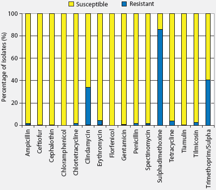
The University of Minnesota Veterinary Diagnostic Laboratory (MN VDL) uses the disk diffusion technique to test for H parasuis antimicrobial susceptibility. In our hands, this technique yields more reliable and reproducible results. Drugs included in the susceptibility panel are ampicillin, ceftiofur, cephalothin, chlortetracycline, clindamycin, erythromycin, florfenicol, gentamicin, penicillin, spectinomycin, sulphadimethoxine, tetracycline, tiamulin, tilmicosin, and trimethoprim-sulphamethoxazole. Updated information on H parasuis antimicrobial susceptibility profiles obtained in the MN VDL in the fiscal year of 2006 is shown in Figure 1. According to this data, H parasuis is susceptible to most antibiotics. However, resistance genes to antibiotics commonly used in swine production have been recently reported. Tetracycline resistance genes, more specifically Tet B, have been found in plasmids recovered from H parasuis isolates involved in an outbreak.8 Beta-lactam resistance genes are currently being characterized.9 The use of genotypic approaches will certainly improve our understanding of antibiotic resistance in H parasuis.
| Figure 1: Haemophilus parasuis antimicrobial
susceptibility profiles obtained at the University of Minnesota Veterinary
Diagnostic Laboratory during the fiscal year of 2006.
|
Interpretation
An isolate is reported as susceptible, intermediate, or resistant to an antibiotic depending on the recommendations of the CLSI for interpretation of results obtained using disk diffusion and microdilution. Occasionally, H parasuis will not grow in the media used for antimicrobial susceptibility testing. In these cases, trends in antibiotic susceptibility profiles can be used for selection of drugs to be used for treatment (Figure 1). Failures in antibiotic treatments may occur even when susceptibility testing indicates that drugs should be effective against a specific isolate. Although disk diffusion and microdilution techniques have limitations, especially when testing fastidious organisms, many other factors, including management practices, route of administration, compliance, and concurrent viral infections, may affect the outcome of antibiotic treatments.
Serotyping
Haemophilus parasuis serotyping provides important
information for selection of commercial vaccines. There is good
protection within serotype groups, whereas cross-protection between
different serotypes is generally poor.1 Two techniques
are available for H parasuis serotyping: the agar gel
precipitation test (AGPT) and indirect hemagglutination
(IHA).10,11 The AGPT was the first technique developed
to serotype H parasuis. This technique uses heat-treated
antigens. Extracts are prepared by autoclaving bacterial
suspensions for 2 hours at 121°C, then centrifuging. The
supernatants are used for serotyping. The AGPT is performed on a
glass slide containing agar gel with wells filled with antigen or
rabbit sera specific for the 15
H parasuis serotypes. Precipitation lines are read after 24
hours of incubation.10
The IHA is also performed using heat-treated antigens. Formalin-inactivated bacterial cells are boiled and centrifuged, and the resulting supernatant is used to coat sheep red blood cells (SRBC). The test is performed using a microtiter system. Serial twofold dilutions of sera are made in saline in U-bottom microplates. Sensitized SRBC suspensions are added to the wells and plates and incubated at 37°C for 2 hours. The IHA titer is expressed as the reciprocal of the highest dilution of serum showing a definite positive pattern (flat sediment) compared with the pattern of negative control (smooth dot in the center of the well).11,12
The literature reports that the AGPT yields a higher percentage of nontypable isolates (15% to 41%) compared with the IHA test (< 10%).1,11
Interpretation
There are 15 known serotypes of H parasuis. Interpretation of serotyping results using AGPT and IHA is straightforward. Nevertheless, analysis of results may be subjective. Some field isolates do not produce enough antigens in vitro to be serotyped. Cross-reaction between serotypes may occur, and results are reported on the basis of the strongest reaction among several positive results. Nontypable isolates may generate cross-reactions that impair accurate serotype allocation. They may also represent new serotypes for which antisera are not available.
Detection by PCR
Isolation of H parasuis from clinical samples is necessary for antimicrobial susceptibility testing, serotyping, and genotyping. However, this fastidious microorganism requires a special medium for growth and survives for a short period of time (8 hours) at room temperature.13 Samples from pigs that are found dead are frequently culture-negative.4 We have recently standardized and validated a PCR test to detect H parasuis in clinical samples (Hps-PCR).14 The Hps-PCR test being offered at the MN VDL is a modification of a test previously published. The new test is specific for detection of H parasuis and was validated using 300 clinical samples submitted to the VDL for routine testing. Fibrin recovered from the surfaces of organs with fibrinous serositis was used for bacterial isolation and PCR testing. Of the 300 samples tested, 146 (48.6%) were positive for H parasuis by PCR, and 37 (12.3%) were positive by isolation. Most PCR-positive and isolation-positive results originated from samples with acute lesions (104 by PCR and 22 by isolation), determined by histopathological evaluation. Polymerase chain reaction was also positive for nine samples with subacute lesions and 20 samples with chronic lesions, compared with three and seven samples, respectively, that were positive by isolation. The PCR test detected H parasuis in samples containing mixed bacteria, such as Actinobacillus suis, A pleuropneumoniae, A indolicus, A minor, A porcinus, Bordetella bronchiseptica, Pasteurella multocida, Escherichia coli, and Streptococcus suis. Testing these bacterial species by the PCR test generated negative results, confirming the laboratory specificity of the test for detection of H parasuis. The PCR test was also successful in detecting H parasuis from samples of acute lesions with negative isolation results. The PCR is far more sensitive than traditional bacterial isolation for diagnosis of H parasuis systemic infection.15
Interpretation
A positive PCR result means that H parasuis DNA was detected in the clinical sample. Diagnosis of H parasuis infection on the basis of a positive PCR result is valid. However, isolation should be pursued for antibiotic susceptibility testing, serotyping, and genotyping. Positive PCR results for samples from the nasal cavity, trachea, and lungs are meaningful only if the herd is negative for H parasuis. Samples to be submitted for PCR testing should include the fibrinous exudate present in the pleura, pericardium, peritoneum, spleen, liver, joints, and meninges. Fibrinous exudate may be collected using a swab. Although gross lesions may be absent in meningitis cases, a swab of the brain surface may still be submitted for bacterial isolation and PCR testing.
Genotyping
Haemophilus parasuis genotyping is performed using the enterobacterial repetitive intergenic consensus-based PCR, also known as ERIC-PCR. This technique has greatly improved our understanding of H parasuis epidemiology. The ERIC-PCR has allowed identification of strain variability within serotype groups, and is thus more suitable for epidemiological studies. It has also exposed the high genetic variability existing among H parasuis field isolates and has helped to identify differences between respiratory and systemic strains.16 This technique is currently used by swine veterinarians to identify the sources of virulent strains introduced into the herd, to detect prevalent groups of strains involved in mortality, and for selection of isolates to be used in autogenous vaccines. It is an important tool for surveillance, prevention, and control of H parasuis infections.
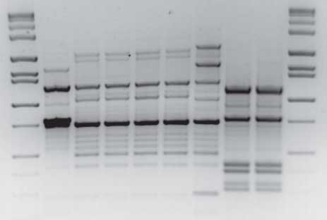
ERIC elements are DNA sequences that are distributed throughout the bacterial genome. These sequences were initially identified in Salmonella species and in Escherichia coli, hence the name “enterobacterial.” They were later found to be highly conserved among different bacterial species. Several copies of ERIC elements sharing the same DNA sequence may be found in a given bacterial genome; therefore, these sequences are referred to as “repetitive” elements. ERIC elements are noncoding regions located between actual genes or coding sequences, so they are distributed between genes, ie, in “intergenic” positions. These elements contain highly conserved central inverted repeats, which are referred to as “consensus.” Their relative positions in the genome of a particular bacterial isolate appear to be conserved in closely related strains. Different strains have different distributions of ERIC elements in their genomes. The PCR amplification of genomic regions between ERIC copies produces a collection of distinct fragments on an agarose gel. These fragments generate a genomic fingerprint, which can be used to identify groups of related strains (Figure 2).1,17
Genomic fingerprints may be compared manually or by computer software. Manual assignment of strain groups is easily performed when a small number of isolates needs to be compared. Computer programs are especially useful to organize large databases of genomic fingerprints in family trees or dendrograms. At the MN VDL, we use the GelCompare software to manage our genomic databases (available at https://mvdl.auxs.umn.edu/vetlabs/genomics.html).
The first step in constructing a genomic fingerprint-based dendrogram is to run the ERIC-PCR and to obtain the picture of the agarose gel containing the DNA fragments to be analyzed (Figure 2). The gel picture is then uploaded into the GelCompare software, and lanes containing genomic fingerprints and molecular size markers are identified. Molecular markers located in the first and last lanes of the gel are used to align or normalize the gel picture. The same marker is added as a “reference” in the database, so each new gel that is analyzed is adjusted to the database reference system. This procedure corrects for small differences between gel runs and allows comparison of genomic fingerprints from different gels. A positive control with a known genomic fingerprint is also added to each PCR reaction to assure reproducibility of the method. This positive control is compared with previous controls in the database, and a PCR reaction is either accepted or rejected by comparing presence, absence, and intensity of bands from positive controls obtained in each run. The amount of DNA used in the PCR reaction is an important source of variation in band intensity. It is very important to use the same amount of extracted DNA for each bacterial isolate so that genomic fingerprints are comparable. We use 100 ng of DNA per bacterial isolate.16
| Figure 2: Agarose gel containing Haemophilus
parasuis genomic fingerprints. Lanes 1 and 10: molecular size markers.
Lanes 2–9: H parasuis genomic fingerprints.
|
Genomic fingerprints may be compared using either a band-based or a curve-based method. The band-based method compares fingerprints by identifying the presence or absence of bands in a specific position. This method does not take into account the intensity of the bands. The curve-based method takes into account not only the presence, absence, and location of each band, but also its intensity. This method has proven to be more reliable than band matching for comparison of H parasuis genomic fingerprints.16 However, it is also more sensitive to small variations in the brightness and contrast of different gel pictures and to variations in results obtained in different PCR runs.18
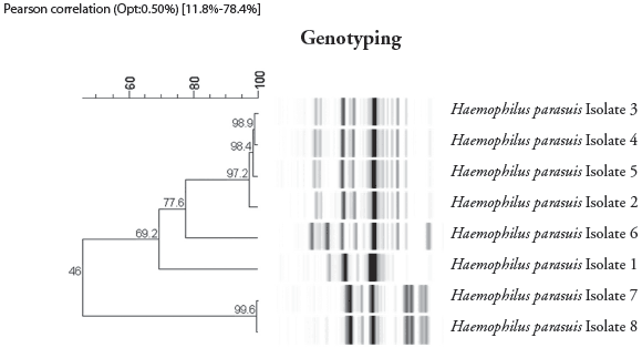
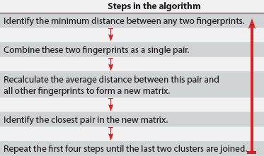
Once the gel picture is standardized and densitometric curves are generated for each band, the next step is to construct a dendrogram. The input of a clustering method is a similarity matrix and the output is a dendrogram. Several mathematical models can be used to generate a similarity matrix. We use the Pearson correlation coefficient to generate similarity matrices based on comparison of densitometric curves. Once the similarity matrices are generated, a clustering method is selected for construction of the dendrogram. A variety of algorithms for hierarchical or divisive clustering analyses generating dendrograms are available.19 We use the unweighted pair-group method, using arithmetic averages (UPGMA) to construct H parasuis dendrograms (Figure 3).16 UPGMA is a straightforward method of tree construction that uses an algorithm shown in Figure 4.
| Figure 3: Construction of a dendrogram for Haemophilus
parasuis using the gel picture shown in Figure 2. The similarity
matrix was calculated using the Pearson correlation coefficient. Cluster
analysis was performed using the unweighted pair-group method with optimization
of 0.5%. Four groups of strains or clusters were identified.
|
| Figure 4: Algorithm for hierarchical clustering
analyses17,19 used to construct dendrograms such as the one
for Haemophilus parasuis shown in Figure 3.
|
Interpretation
The definition of strain is controversial, and may be considerably different depending on the infectious agent being evaluated. For H parasuis, isolates sharing the same genomic fingerprint (identical band pattern, including location and intensity of bands) are considered the same strain. A genomic fingerprint-based dendrogram is different from a sequence-based dendrogram. Dendrograms constructed on the basis of data retrieved from gel pictures are sensitive to small differences in mobility of bands in the agar gel and brightness and contrast of the pictures used for analysis. Even when laboratory conditions are strictly standardized, it is rare to produce genomic fingerprints that are 100% similar to each other. The dendrograms are very useful to organize multiple genomic fingerprints in clusters of closely related strains. However, manual inspection of band patterns, including presence, absence, and intensity, is still important for accurate identification of different strains.
Genotype and serotype results are usually associated, meaning that isolates from a strain group may have the same serotype. Haemophilus parasuis isolates sharing similar genomic fingerprints may occasionally have different serotypes.16 These differences may be real or they may be the result of subjective interpretation of serotyping techniques.
Haemophilus parasuis isolates with similar genomic fingerprints may have different antibiotic resistance profiles. As for serotyping, these differences may be associated with the technique used for susceptibility testing or they may be the result of conditions used in different laboratories. Different resistance profiles may also be associated with the presence of resistance genes in extragenomic elements, eg, plasmids.
Summary
Five techniques are currently available for H parasuis diagnosis: bacterial isolation, antimicrobial susceptibility testing, serotyping, detection by PCR, and genotyping. These techniques provide unique and complementary diagnostic information that can be used for surveillance, prevention, and control of H parasuis. Detection of H parasuis in clinical samples by PCR can be used to define the role of this pathogen in mortality, especially when bacterial isolation is negative. However, H parasuis isolation is still necessary for antibiotic susceptibility testing, serotyping, and genotyping. Antimicrobial susceptibility profiles may vary between laboratories, especially if different techniques are used for testing. Serotyping can be performed using AGPT and IHA. Although IHA is reportedly more sensitive than AGPT, nontypable isolates are still obtained using both techniques. Genotyping is an excellent epidemiological tool that can be used for surveillance and control of H parasuis infections. Epidemiological studies are more robust if information from isolation site, serotyping, genotyping, and antimicrobial susceptibility profiles are analyzed in combination.
References
1. Oliveira S, Pijoan C. Haemophilus parasuis: new trends on diagnosis, epidemiology and control. Vet Microbiol. 2004;99:1–12.
2. Moller K, Fussing V, Grimont PA, Paster BJ, Dewhirst FE, Kilian M. Actinobacillus minor sp. nov., Actinobacillus porcinus sp. nov., and Actinobacillus indolicus sp. nov., three new V factor-dependent species from the respiratory tract of pigs. Int J Syst Bacteriol. 1996;46:951–956.
3. Kielstein P, Wuthe H, Angen O, Mutters R, Ahrens P. Phenotypic and genetic characterization of NAD-dependent Pasteurellaceae from the respiratory tract of pigs and their possible pathogenetic importance. Vet Microbiol. 2001;81:243–255.
*4. Oliveira S. Improving rate of success in isolating Haemophilus parasuis from clinical samples. J Swine Health Prod. 2004;12:308–309.
5. NCCLS. Performance Standards for Antimicrobial Disk and Dilution Susceptibility Tests for Bacteria Isolated from Animals; Approved Standard. 2nd ed. NCCLS document M31-A2 [ISBN 1–56238–461–9]. NCCLS, 940 West Valley Road, Suite 1400, Wayne, PA 19087–1898; 2002.
6. Clinical and Laboratory Standards Institute. Methods for Antimicrobial Dilution and Disk Susceptibility Testing of Infrequently Isolated or Fastidious Bacteria; Proposed Guideline. CLSI document M45-P [ISBN 1–56238–583–6]. Clinical and Laboratory Standards Institute, 940 West Valley Road, Suite 1400, Wayne, PA 19087–1898; 2005.
7. Clinical and Laboratory Standards Institute. Methods for Dilution Antimicrobial Susceptibility Tests for Bacteria That Grow Aerobically; Approved Standard. 7th ed. Clinical and Laboratory Standards Institute document M7-A7 [ISBN 1–56238–587–9]. Clinical and Laboratory Standards Institute, 940 West Valley Road, Suite 1400, Wayne, PA 19087–1898; 2006.
8. Lancashire JF, Terry TD, Blackall PJ, Jennings MP. Plasmid-encoded Tet B tetracycline resistance in Haemophilus parasuis. Antimicrob Agents Chemother. 2005;49:1927–1931.
9. San Millan A, Escudero JA, Catalan AM, Porrero MC, Dominguez L, Moreno MA, Gonzalez-Zorn B. R1940 Beta-lactam resistance in Haemophilus parasuis. Clin Microbiol Infect. 2006;12(suppl 4):1.
10. Morozumi T, Nicolet J. Some antigenic properties of Haemophilus parasuis and a proposal for serological classification. J Clin Microbiol. 1986;23:1022–1025.
11. Tadjine M, Mittal KR, Bourdon D, Gottschalk M. Development of a new serological test for serotyping Haemophilus parasuis isolates and determination of their prevalence in North America. J Clin Microbiol. 2004;42:839–840.
12. Mittal KR, Higgins R, Lariviere S. Determination of antigenic specificity and relationship among Haemophilus pleuropneumoniae serotypes by an indirect hemagglutination test. J Clin Microbiol. 1983;17:787–790.
13. Morozumi T, Hiramune T. Effect of temperature on the survival of Haemophilus parasuis in physiological saline. Natl Inst Anim Health Q (Tokyo). 1982;22:90–91.
14. Oliveira S, Galina L, Pijoan C. Development of a PCR test to diagnose Haemophilus parasuis infections. J Vet Diag Invest. 2001;13:495–501.
*15. Oliveira S, Tomasezewski J, Gayle R, Collins J. Validation of a PCR test for detection of Haemophilus parasuis in clinical samples. Proc 49th AAVLD Ann Meet. 2006:134.
16. Oliveira S, Blackall PJ, Pijoan C. Characterization of the diversity of Haemophilus parasuis field isolates by use of serotyping and genotyping. Am J Vet Res. 2003;64:435–442.
17. Versalovic J, Koeuth T, Lupski JR. Distribution of repetitive DNA sequences in eubacteria and application to fingerprinting of bacterial genomes. Nucleic Acids Res. 1991;9:6823–6831.
*18. Oliveira S, Oleson T, Titus M, Simonson R. Comparison of Haemophilus parasuis genotyping using ERIC-PCR and AFLP. Proc AASV. Des Moines, Iowa. 2004:273–276.
19. van Ooyen A. Theoretical aspects of pattern analysis. In: Dijkshoorn L, Tower KJ, Struelens M, eds. New Approaches for the Generation and Analysis of Microbial Fingerprints. Amsterdam, The Netherlands: Elsevier; 2001:31–45.
* Non-refereed references.


