Brief communication |
Peer reviewed |
Fecal shedding of a highly cell-culture-adapted porcine epidemic diarrhea virus after oral inoculation in pigs
Excreción fecal de un virus de diarrea epidémica porcina altamente adaptado al cultivo celular después de la inoculación oral en cerdos
Excrétion fécale d’un virus de la diarrhée épidémique porcin très adapté au culture cellulaire après de l’inoculation orale dans les porcs
DaeSub Song, DVM, PhD; JinSik Oh, DVM, MS, PhD; BoKyu Kang, DVM; JeongSun Yang, DVM; JuYoung Song, DVM; HyoungJoon Moon, DVM; TaeYung Kim, DVM, MS; HanSang Yoo, DVM, MS, PhD; YongSuk Jang, MS, PhD; BongKyun Park, DVM, MS, PhD
DSS, JSO, BKK, JSY, JYS, HJM, TYK, HSY, BKP: Department of Veterinary Medicine and the Xenotransplantation Research Center, College of Veterinary Medicine and School of Agricultural Biotechnology, Seoul National University, Seoul 151-742, Korea. YSJ: Division of Biological Science, the Institute for Molecular Biology and Genetics, Chonbuk National University, Chonju 561-756, Korea. Corresponding author: Dr BongKyun Park, Department of Veterinary Medicine Virology Laboratory, College of Veterinary Medicine, Seoul National University, Seoul 151-742, Korea; Tel: +82-2-880-1255; Fax: +82-2-885-0263; E-mail: parkx026@snu.ac.kr
Cite as: Song DS, Oh JS, Kang BK, et al. Fecal shedding of a highly cell-culture-adapted porcine epidemic diarrhea virus after oral inoculation in pigs. J Swine Health Prod. 2005;13(5):269-272.
Also available as a PDF.
SummaryAfter oral inoculation with a highly cell-culture-adapted strain of porcine epidemic diarrhea virus, shedding was detected by reverse transcription-polymerase chain reaction and restriction fragment length polymorphism for up to 6 days in 3-day-old piglets, 9 days in 2-week-old pigs, and 3 days in late-term pregnant sows. | ResumenDespués de la inoculación oral con una cepa altamente adaptada al cultivo celular del virus de la diarrea epidémica porcina, se detectó excreción por medio de la trascripción inversa de la reacción en cadena de la polimerasa y el polimorfismo de restricción de longitud fragmentaria hasta por 6 días en lechones de 3 días de edad, 9 días en cerdos de 2 semanas de edad y 3 días en hembras gestantes al final del periodo. | ResuméAprès de l’inoculation orale avec une souche très adapté au culture cellulaire du virus de la diarrhée épidémique porcin, l’excrétion a été détecté par la transcription inverse de la réaction en chaîne du polymérase et le polymorphie de restriction de la longueur du fragment pour 6 jours dans porcelets de 3 jours, 9 jours dans les porcelets de 2 semaines, et 3 jours dans les truies gestants à la fin du gestation. |
Keywords: swine, porcine
epidemic
diarrhea virus, oral inoculation, fecal shedding
Search the AASV web site
for pages with similar keywords.
Received: October
28, 2004
Accepted: January
26, 2005
Porcine epidemic diarrhea virus (PEDV), an enveloped, single- stranded RNA virus1,2 in the family Coronaviridae, causes severe diarrhea in swine. The main and only obvious clinical sign of porcine epidemic diarrhea (PED) is watery diarrhea. Mortality may be as high as 80%, but averages 50%.3,4 Piglets up to 1 week of age may die from dehydration after 3 to 4 days. In Europe, weaned pigs and even adult pigs have been severely affected, while disease in suckling pigs in the same herds was mild, even in the absence of immunity.5 The virus is transmitted in feces from infected pigs.6 Detection of PEDV antigen in feces after inoculation varies with virulence and inoculated dose.7 In a previous study, PEDV strain DR13 was isolated and serially passaged in Vero cell cultures.8 The highly adapted virus was differentiated from wild-type viruses by reverse transcriptase-polymerase chain reaction (RT-PCR) and restriction fragment length polymorphism (RFLP).8,9
The objective of this study was to describe fecal shedding of PEDV strain DR13, detected by RT-PCR and RFLP, after oral inoculation of piglets and older pigs. Preliminary work showed that when 3-day-old piglets were inoculated with the parent strain (ie, non-attenuated DR13), mortality was high and time between inoculation and death was short. Therefore, 2-week-old pigs were the main focus of the shedding study in young animals.
Materials and methods
Virus propagation
Strain DR13 (PEDV parent strain) was harvested from a suspension of whole minced small intestine from infected neonatal pigs. A continuous Vero cell line (ATCC CCL-81) was maintained in [alpha]-minimum essential medium (MEM) supplemented with 5% fetal bovine serum, penicillin (100 units per mL), streptomycin (100 mg per mL), and amphotericin B (0.25 mg per mL). The cell-attenuated strain DR13 was propagated as previously described.10,11
Oral inoculation and fecal sampling
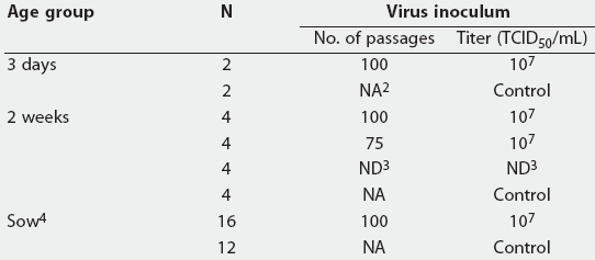
Experiment One. A total of four 3-day-old piglets (Yorkshire x Landrace x Duroc) that were PEDV-seronegative (serum neutralization12 titer 1:<= 2) were employed to investigate fecal shedding after inoculation with cell-culture-adapted (100 passages) strain DR13 (P100; Table 1). The sham-inoculated controls were housed separately. Rectal swabs were collected daily (three cotton-tipped swabs per pig) until 10 days postinoculation (PI).
Table 1: Experiment design for oral inoculation of porcine epidemic diarrhea virus1 strain DR13 in pigs of different ages
1 All 3-day and 2-week-old piglets were inoculated with 5 mL of virus suspension, and sows with 1 mL. Strain DR13 was harvested from intestinal mucosa of infected pigs and either administered as a 10% suspension (parent strain; 0 passages) or propagated in Vero cells, with virus titers expressed as median tissue culture infectious doses (TCID50) per mL. Controls were sham-inoculated with 5 mL (piglets) or 1 mL (sows) of cell culture minimal essential medium. 2 NA = not applicable. 3 ND = not done. Virus titer of the parent strain not determined. 4 Pregnant sows 4 to 5 weeks before farrowing. |
Experiment Two. A total of 16 healthy, 2-week-old mixed-breed pigs were randomly divided into four treatment groups (Table 1). Groups were orally inoculated with strain DR13 either unpassaged (Parent) or passaged 75 (P75) or 100 times (P100), or were sham-inoculated (Control). Rectal swabs were collected daily until 10 days PI.
Experiment Three. Strain DR13 (P100) was tested as described (Table 1) in a total of 16 Yorkshire x Landrace sows in four commercial swine herds (using five of the 500 sows in Herd A; five of the 300 sows in Herd B; three of the 1300 sows in Herd C; and three of the 1200 sows in Herd D). Before inoculation, Herds A and B were PCR-negative for PEDV, and Herds C and D were PCR-positive. In each herd, an equal number of controls were sham-inoculated. Three rectal swabs per sow were collected daily until 5 days PI.
Housing and animal care
Three-day-old piglets and 2-week-old pigs were housed at a university facility in environmentally controlled nurseries with slatted metal flooring and mechanical ventilation. One self feeder and one nipple waterer were provided in each 1.3-m x 1.3-m (1.69 m2) pen. Piglets were housed two per pen and were fed commercial milk replacer. Two-week-old pigs were housed four per pen with ad libitum access to feed and water.
Sows were housed individually in gestation stalls in modern commercial swine facilities. Sows had free access to water and were fed three times a day.
All animal experiments complied with the current laws of Korea. Care and treatment of animals were conducted in accordance with protocols and guidelines of the Seoul National University Institutional Animal Care and Use Committee.
Detection of PEDV and differentiation of cell-culture-adapted PEDV from field isolates
Three cotton swabs were resuspended in 1 mL of MEM. Tubes were vortexed, and the suspension clarified by centrifugation for 10 minutes at 4800g to eliminate large fecal debris.9 Reverse transcription using reverse primer and RT-PCR9 and differentiation of PEDVs8 were performed as previously described.
Results
Experiment One. Porcine epidemic diarrhea virus was detected by RT-PCR in fecal samples from inoculated piglets for 6 days PI, but never in samples from control pigs.
Experiment Two. Fecal samples from inoculated pigs were RT-PCR-positive for PEDV for 8 days PI in the P75 group, 7 days PI in the P100 group, and 9 days PI in the Parent group. No pigs were positive on the day of inoculation or on day 10 PI, and Controls were never positive.
Experiment Three. In Herd A, PEDV was detected in fecal samples from four of five inoculated sows on day 2 PI, and in one sow on day 3 PI. In Herd B, PEDV was detected in fecal samples from all five sows 1 and 2 days PI, and from two of these sows on day 3 PI. In both Herds A and B, detected virus had the same RFLP pattern as that of the inoculum.
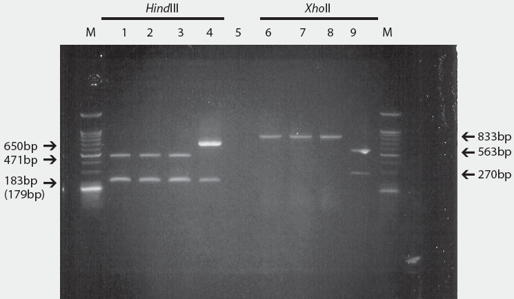
In Herd C, wild-type PEDV was detected in fecal samples from one sow on the day of inoculation, but not thereafter. Fecal samples from that sow and the two others were PCR-positive for the cell-adapted strain DR13 on days 1 and 2 PI. The RFLP patterns of the wild-type PEDV and the cell-adapted strain DR13 (100 passages) are shown in Figure 1.
Figure 1: Restriction fragment length polymorphism patterns of porcine epidemic diarrhea virus (PEDV) isolates and cell-adapted PEDV strain DR13 using restriction enzymes, HindIII and XhoII. From left to right: Lane M, 100 bp DNA ladder; Lanes 1 and 6, KPEDV-9, a strain used in commercial vaccines in South Korea; Lanes 2 and 7, wild type PEDV isolate B409; Lanes 3 and 8, parent strain DR13; Lanes 4 and 9, cell-adapted strain DR13 (passage level 100); Lane 5, control.
|
In Herd D, wild-type PEDV was detected in fecal samples from all three sows on the day of inoculation, and one of these sows continued to shed only the wild-type strain on days 1 through 5 PI. The other two sows were PCR-positive for both the wild-type and cell-adapted strains until days 2 and 3 PI, respectively.
All fecal samples from controls in all four herds were PCR-negative for PEDV at all collection points. No pigs died during this study.
Discussion
In this study, it was possible to monitor fecal shedding of PEDV under commercial conditions because the inoculating strain of PEDV could be differentiated from wild-type PEDV. The short shedding period in sows might have been associated with age dependency, interference by PEDV antibody, or virus attenuation. In Herd D, the two sows that shed both the cell-adapted and wild-type PEDV might have been co-infected with wild virus after oral inoculation with the cell-culture-adapted strain DR13. The third sow in Herd D, that shed only wild-type PEDV, might have been infected with wild-type virus before inoculation, ie, few susceptible enterocytes were available for replication of the cell-culture-adapted PEDV. Alternatively, errors in oral inoculation, fecal sampling, or virus detection may have occurred in this sow.
In another study,5 PEDV antigens were detected in fecal samples from 3 until 11 days PI, with peak excretion at 4 and 5 days. Failure to detect viral antigen in fecal samples from pigs inoculated with attenuated PEDV has been previously reported,13,14 and pigs infected with virulent virus shed more virus for longer periods than pigs inoculated with cell-culture-adapted virus.8 However, in gnotobiotic pigs, replication of attenuated virus occurred at any dose.15 These inconsistencies might be due to differences in method of virus detection technique (eg, ELISA or RT-PCR). Detection of PEDV in feces might be affected by the reliability and sensitivity of the technique.16 Detection of viral antigen in feces after inoculation of PEDV was markedly influenced by the virulence and the inoculation dose of PEDV.7 Using the RT-PCR technique, virus was detected from clinical samples containing 107 TCID50 per mL.9 Age, breed, nutrition, or fecal sampling might also affect fecal shedding of PEDV, and the normal intestinal microflora may interfere with replication of cell-culture-adapted PEDV.7
In this study, changes in RFLP patterns were identified in PEDV strain DR13 after 75 passages; therefore, strain DR13 attenuated to passage level 75 was used as inoculum to identify the relationship between pathogenicity and RFLP-marker change. However, there was no apparent difference in virus shedding when strain DR13 passaged either 75 or 100 times was inoculated into 2-week-old pigs. In addition, the RFLP marker of the attenuated strain DR13 was maintained during pig passages. Attenuation and immunogenicity of PEDV strain DR13 (passage level 100) have been reported previously.8 This cell-attenuated DR13 could be used as a marker vaccine that can be differentiated from other strains of PEDV.
Implications
- Under the conditions of this study, Vero cell-adapted PEDV strain DR13 can be differentiated from wild-type PEDVs using RT-PCR and RFLP technology.
- Cell-adapted strain DR13 has potential as a PEDV marker vaccine.
Acknowledgements
This work was supported by the Korea Research Foundation Grant (KRF-2002-070-C00069), the Biogreen 21, and the Brain Korea 21 Project of Ministry of Education and Human Resources Development, Republic of Korea.
References
1. Debouck P, Pensaert M. Experimental infection of pigs with a new porcine enteric coronavirus CV777. Am J Vet Res. 1980;41:219-223.
2. Ducatelle R, Coussement W, Pensaert M, Debouck P, Hoorens J. In vivo morphogenesis of a new porcine enteric coronavirus, CV777. Arch Virol. 1981;68:35-44.
3. Wood EN. An apparently new syndrome of porcine epidemic diarrhea. Vet Rec. 1977; 100:243-244.
4. Wood EN. Transmissible gastroenteritis and epidemic diarrhea of pigs. Br Vet J. 1979;135:305-314.
5. Pensaert MB. Porcine epidemic diarrhea virus. In: Straw BE, D’Allaire S, Mengeling WL, Taylor DJ, eds. Diseases of Swine. 8th ed. Ames, Iowa: Iowa State University Press. 1999;179-185.
6. Pensaert MB, Debouck P. A new coronavirus-like agent associated with diarrhea in swine. Arch Virol. 1978;58:243-247.
7. de Arriba ML, Carvajal A, Pozo J, Rubio P. Mucosal and systemic isotype-specific antibody responses and protection in conventional pigs exposed to virulent or attenuated porcine epidemic diarrhea virus. Vet Immunol Immunopathol. 2002;85:85-97.
8. Song DS, Yang JS, Oh JS, Han JH, Park BK. Differentiation of a Vero cell adapted porcine epidemic diarrhea virus from Korean field strains by restriction fragment length polymorphism analysis of ORF 3. Vaccine. 2003;21:1833-1842.
9. Kim SY, Song DS, Park BK. Differential detection of transmissible gastroenteritis virus and porcine epidemic diarrhea virus by duplex RT-PCR. J Vet Diagn Invest. 2001;13:516-520.
10. Hofmann M, Wyler R. Propagation of the virus of porcine epidemic diarrhea in cell culture. J Clin Microbiol. 1988;26:2235-2239.
11. Kwon CH, Kwon BJ, Lee JG, Kwon GO, Kang YB. Derivation of attenuated porcine epidemic diarrhea virus (PEDV) as vaccine candidate. Vaccine. 1999;17:2546-2553.
12. Kusanagi K, Kuwahara H, Katoh T, Nunoya T, Ishikawa Y, Samejima T, Tajima M. Isolation and serial propagation of porcine epidemic diarrhea virus infection in cell cultures and partial characterization of the isolate. J Vet Med Sci. 1992;54:303-318
*13. Carvajal A, Diego R, Lanza I, Rubio P, Carmenes P. Immunobiology of viral infection. Evaluation of an ELISA for the detection of porcine epidemic diarrhea virus (PEDV) in feces of naturally infected pigs. Proc Congr Eur Soc Vet Virol. Interlaken, Switzerland. 1994;3:516-519.
14. Frederick GT, Bohl EH, Cross RF. Pathogenicity of an attenuated strain of transmissible gastroenteritis virus for newborn pigs. Am J Vet Res. 1976;37:165-169.
*15. Bernasconi C, Guscetti F, Utiger A, Van Reeth K, Ackermann M, Pospischil A. Experimental infection of gnotobiotic piglets with a cell culture adapted porcine epidemic diarrhea virus: clinical, histopathological and immunohistochemical findings. Proc Cong Eur Soc Vet Virol. Interlaken, Switzerland. 1994;3:542-546.
16. Guscetti F, Bernasconi C, Tobler K, Van Reeth K, Pospischil A, Ackerman M. Immunohistochemical detection of porcine epidemic diarrhea virus compared to other method. Clin Diagn Lab Immunol. 1998;5:412-414.
* Non-refereed references.

