Practice Tip |
Non refereed |
Transabdominal imagery of ovarian activity using real-time ultrasound
William L. Hollis, DVM
Carthage Veterinary Service Ltd, 34 Main St, Carthage, IL 62321; E-mail: hollis@hogvet.com.
Hollis WL. Transabdominal imagery of ovarian activity using real-time ultrasound. J Swine Health Prod. 2003;11(1):37-38.
Swine veterinary practitioners have implemented on-farm use of real-time ultrasound (RTU) diagnostics for many production systems, and employees have learned to make clear and consistent pregnancy diagnosis using this valuable tool. Swine farms have begun to purchase ultrasound machines, and with daily on-farm availability, experimentation with this technology has increased.
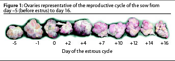
We are using RTU to differentiate normal and abnormal anatomy, and to select appropriate therapy and make culling decisions. Knowledge of the normal reproductive anatomy and reproductive cycle of the sow is necessary to interpret ovarian activity (Figure 1), and training is required to make full use of ultrasound equipment.
 Visualization of the ovary can be achieved transabdominally by using heavy
lubrication and pointing the probe toward the bladder – a full bladder is helpful. Once
the bladder is identified, slowly trace the outline of its dorsal surface cranially.
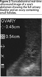
When only very small follicles are present, it is difficult to distinguish the ovary from
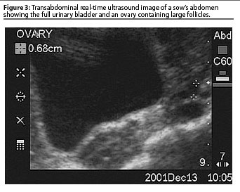
surrounding connective tissue (Figure 2). Large follicles appear like a cluster
of grapes along the cranial edge of the bladder (Figure 3). Once the ovary is
visualized, freeze the image and measure the
diameter of follicles. Researchers tell us a 6-mm
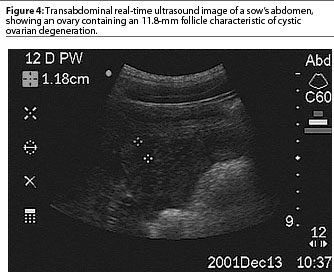
follicle is near ovulation, and any follicle greater than 12 mm is a sign of cystic
ovar-ian degeneration (COD), as shown in Figure 4. A barn staff member or the herd
veterinarian may use RTU for a wide range of diagnostic applications in sows.
Visualization of the ovary can be achieved transabdominally by using heavy
lubrication and pointing the probe toward the bladder – a full bladder is helpful. Once
the bladder is identified, slowly trace the outline of its dorsal surface cranially.
When only very small follicles are present, it is difficult to distinguish the ovary from
surrounding connective tissue (Figure 2). Large follicles appear like a cluster
of grapes along the cranial edge of the bladder (Figure 3). Once the ovary is
visualized, freeze the image and measure the
diameter of follicles. Researchers tell us a 6-mm
follicle is near ovulation, and any follicle greater than 12 mm is a sign of cystic
ovar-ian degeneration (COD), as shown in Figure 4. A barn staff member or the herd
veterinarian may use RTU for a wide range of diagnostic applications in sows.
Real-time ultrasonography is most helpful in the following situations: detection of cystic follicles in non-cyclic weaned sows; detection of ovarian activity in sows diagnosed not pregnant after a normal cycle; monitoring cyclic activity in gilt groups; and prediction of ovulation in relation to split-weaning and early weaning.
Ovarian activity may be delayed in non-cyclic weaned sows for many reasons. Evaluation of cull sows at slaughter has identified COD in up to 10% of sows culled for infertility.1 Most breeding barn production staff are good at making predictions of cycle timing by lactation length. Sows that do not cycle are often pooled for further evaluation. This "late-wean" pool may be evaluated weekly using the RTU. As a full bladder certainly helps to locate the ovaries, perform the examination early in the day if possible.
Gilt groups often contain a small percentage of apparently non-cyclic gilts. These may be injected with PG-600 (Intervet, Millsboro, Delaware) in an effort to bring them into the following weekly group. However, gilts with ovarian follicular activity should not be treated with PG-600. Use of RTU allows the operator to identify ovarianactivity in gilts presumed to be non-cycling.
We most commonly use RTU to visualize the ovary in sows 14 to 21 days after a negative pregnancy check. Females that have not cycled after 21 days of observation must be evaluated for ovarian activity, and ovarian inactivity is grounds for immediate culling. Detecting ovarian activity tells the breeding department leader that observation of the pregnancy-check-negative "opportunity" group has failed to identify potential breeders.
Ovulation timing is a precise measurement that we do not try to achieve with on-farm weekly RTU examinations. We have, however, used RTU to determine if farms with excessive semen use can skip mating at the first identified estrus. Split-weaned sows tend to cycle earlier in the weekly breeding period, and early weaned sows tend to require a delay or even a skip in the breeding period. Both of these groups of females may be evaluated by RTU to determine their impact on the weekly breeding schedule. Evaluation of these females in each breed group alone may influence breeding patterns.
Reviews of on-farm use of RTU are available. Dr Rob Knox, University of Illinois, has provided an excellent article on this topic.2 We have found the SonoSite 180 (SonoSite, Inc, Bothell, Washington) to provide the clearest picture of the bladder and the cranial-dorsal position of the ovar-ian tissue. The Tringa 50s (Phar Vision Ultrasound, Tequesta, Florida) and the Real McCoy (EI Medical, Loveland, Colorado) are also used on sow farms that we manage. All have advantages and disadvantages. Continued use of RTU sharpens the accuracy and speed of the operator and adds value to the machine.
References – refereed
1. Britt JH, Almond GW, Flowers WL. Diseases of the reproductive system. In: Straw BE, D’Allaire S, Mengeling WL, Taylor DJ, eds. Diseases of Swine. 8th ed. Ames, Iowa: Iowa State University Press. 1999:898.
2. Knox RV, Althouse GC. Visualizing the reproductive tract of the female pig using real-time ultrasonography. Swine Health Prod. 1999;7:207-215.